3SOM

crystal structure of human MMACHC


Experimental Data Snapshot

  • Method: X-RAY DIFFRACTION
  • Resolution: 2.40 Å
  • R-Value Free: 0.206 
  • R-Value Work: 0.182 
  • R-Value Observed: 0.183 

wwPDB Validation   3D Report Full Report


Ligand Structure Quality Assessment 


This is version 1.2 of the entry. See complete history


Literature

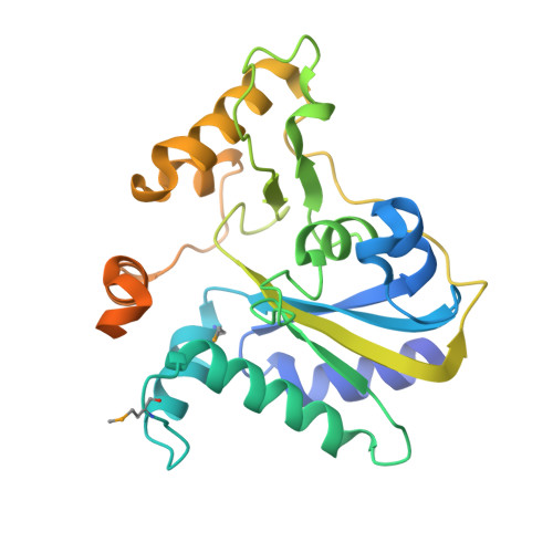
Structure of MMACHC reveals an arginine-rich pocket and a domain-swapped dimer for its B12 processing function.

Froese, D.S.Krojer, T.Wu, X.Shrestha, R.Kiyani, W.von Delft, F.Gravel, R.A.Oppermann, U.Yue, W.W.

(2012) Biochemistry 51: 5083-5090

  • DOI: https://doi.org/10.1021/bi300150y
  • Primary Citation of Related Structures:  
    3SOM

  • PubMed Abstract: 

    Defects in the MMACHC gene represent the most common disorder of cobalamin (Cbl) metabolism, affecting synthesis of the enzyme cofactors adenosyl-Cbl and methyl-Cbl. The encoded MMACHC protein binds intracellular Cbl derivatives with different upper axial ligands and exhibits flavin mononucleotide (FMN)-dependent decyanase activity toward cyano-Cbl as well as glutathione (GSH)-dependent dealkylase activity toward alkyl-Cbls. We determined the structure of human MMACHC·adenosyl-Cbl complex, revealing a tailor-made nitroreductase scaffold which binds adenosyl-Cbl in a "base-off, five-coordinate" configuration for catalysis. We further identified an arginine-rich pocket close to the Cbl binding site responsible for GSH binding and dealkylation activity. Mutation of these highly conserved arginines, including a replication of the prevalent MMACHC missense mutation, Arg161Gln, disrupts GSH binding and dealkylation. We further showed that two Cbl-binding monomers dimerize to mediate the reciprocal exchange of a conserved "PNRRP" loop from both subunits, serving as a protein cap for the upper axial ligand in trans and required for proper dealkylation activity. Our dimeric structure is supported by solution studies, where dimerization is triggered upon binding its substrate adenosyl-Cbl or cofactor FMN. Together our data provide a structural framework to understanding catalytic function and disease mechanism for this multifunctional enzyme.


  • Organizational Affiliation

    Structural Genomics Consortium, NIHR Oxford Biomedical Research Unit, University of Oxford, Oxford, U.K.


Macromolecules
Find similar proteins by:  (by identity cutoff)  |  3D Structure
Entity ID: 1
MoleculeChains Sequence LengthOrganismDetailsImage
Methylmalonic aciduria and homocystinuria type C protein
A, B, C, D, E
A, B, C, D, E, F, G, H, I, J, K, L, M, N, O, P
286Homo sapiensMutation(s): 0 
Gene Names: MMACHC
UniProt & NIH Common Fund Data Resources
Find proteins for Q9Y4U1 (Homo sapiens)
Explore Q9Y4U1 
Go to UniProtKB:  Q9Y4U1
PHAROS:  Q9Y4U1
GTEx:  ENSG00000132763 
Entity Groups  
Sequence Clusters30% Identity50% Identity70% Identity90% Identity95% Identity100% Identity
UniProt GroupQ9Y4U1
Sequence Annotations
Expand
  • Reference Sequence
Small Molecules
Ligands 5 Unique
IDChains Name / Formula / InChI Key2D Diagram3D Interactions
B12
Query on B12

Download Ideal Coordinates CCD File 
AC [auth P]
CA [auth D]
CB [auth J]
FA [auth E]
FB [auth K]
AC [auth P],
CA [auth D],
CB [auth J],
FA [auth E],
FB [auth K],
KA [auth F],
KB [auth L],
NA [auth G],
NB [auth M],
Q [auth A],
SB [auth N],
TA [auth H],
V [auth B],
VB [auth O],
WA [auth I],
X [auth C]
COBALAMIN
C62 H89 Co N13 O14 P
LKVIQTCSMMVGFU-DWSMJLPVSA-N
5AD
Query on 5AD

Download Ideal Coordinates CCD File 
AA [auth C]
AB [auth I]
BA [auth D]
BB [auth J]
HA [auth E]
AA [auth C],
AB [auth I],
BA [auth D],
BB [auth J],
HA [auth E],
IB [auth K],
JA [auth F],
JB [auth L],
QA [auth G],
QB [auth M],
RA [auth G],
RB [auth N],
SA [auth H],
T [auth A],
U [auth B],
YB [auth O],
ZA [auth I],
ZB [auth P]
5'-DEOXYADENOSINE
C10 H13 N5 O3
XGYIMTFOTBMPFP-KQYNXXCUSA-N
FLC
Query on FLC

Download Ideal Coordinates CCD File 
BC [auth P]
DA [auth D]
DB [auth J]
GA [auth E]
GB [auth K]
BC [auth P],
DA [auth D],
DB [auth J],
GA [auth E],
GB [auth K],
LA [auth F],
LB [auth L],
OA [auth G],
OB [auth M],
R [auth A],
TB [auth N],
UA [auth H],
W [auth B],
WB [auth O],
XA [auth I],
Y [auth C]
CITRATE ANION
C6 H5 O7
KRKNYBCHXYNGOX-UHFFFAOYSA-K
DHL
Query on DHL

Download Ideal Coordinates CCD File 
CC [auth P]
EA [auth D]
EB [auth J]
HB [auth K]
MA [auth F]
CC [auth P],
EA [auth D],
EB [auth J],
HB [auth K],
MA [auth F],
MB [auth L],
PA [auth G],
PB [auth M],
S [auth A],
UB [auth N],
VA [auth H],
XB [auth O],
YA [auth I],
Z [auth C]
2-AMINO-ETHANETHIOL
C2 H7 N S
UFULAYFCSOUIOV-UHFFFAOYSA-N
EDO
Query on EDO

Download Ideal Coordinates CCD File 
DC [auth P],
EC [auth P],
IA [auth E]
1,2-ETHANEDIOL
C2 H6 O2
LYCAIKOWRPUZTN-UHFFFAOYSA-N
Modified Residues  1 Unique
IDChains TypeFormula2D DiagramParent
MSE
Query on MSE
A, B, C, D, E
A, B, C, D, E, F, G, H, I, J, K, L, M, N, O, P
L-PEPTIDE LINKINGC5 H11 N O2 SeMET
Experimental Data & Validation

Experimental Data

  • Method: X-RAY DIFFRACTION
  • Resolution: 2.40 Å
  • R-Value Free: 0.206 
  • R-Value Work: 0.182 
  • R-Value Observed: 0.183 
  • Space Group: P 1
Unit Cell:
Length ( Å )Angle ( ˚ )
a = 71.664α = 88.53
b = 71.96β = 85.25
c = 300.055γ = 83.76
Software Package:
Software NamePurpose
GDAdata collection
SHARPphasing
BUSTERrefinement
XDSdata reduction
SCALAdata scaling

Structure Validation

View Full Validation Report


Ligand Structure Quality Assessment 


Entry History 

Revision History  (Full details and data files)

  • Version 1.0: 2011-07-27
    Type: Initial release
  • Version 1.1: 2012-10-24
    Changes: Non-polymer description
  • Version 1.2: 2015-04-15
    Changes: Database references