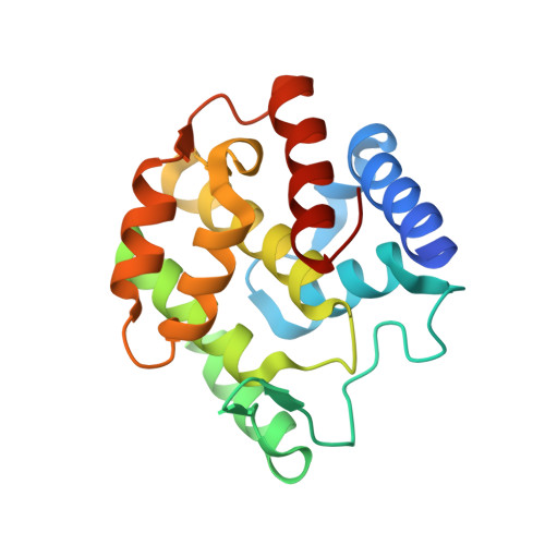
Adenylylation control by intra- or intermolecular active-site obstruction in Fic proteins.
Engel, P., Goepfert, A., Stanger, F.V., Harms, A., Schmidt, A., Schirmer, T., Dehio, C.(2012) Nature 482: 107-110
- PubMed: 22266942
- DOI: https://doi.org/10.1038/nature10729
- Primary Citation of Related Structures:
3S6A, 3SE5, 3SHG, 3SN9 - PubMed Abstract:
Fic proteins that are defined by the ubiquitous FIC (filamentation induced by cyclic AMP) domain are known to catalyse adenylylation (also called AMPylation); that is, the transfer of AMP onto a target protein. In mammalian cells, adenylylation of small GTPases through Fic proteins injected by pathogenic bacteria can cause collapse of the actin cytoskeleton and cell death. It is unknown how this potentially deleterious adenylylation activity is regulated in the widespread Fic proteins that are found in all domains of life and that are thought to have critical roles in intrinsic signalling processes. Here we show that FIC-domain-mediated adenylylation is controlled by a conserved mechanism of ATP-binding-site obstruction that involves an inhibitory α-helix (α(inh)) with a conserved (S/T)XXXE(G/N) motif, and that in this mechanism the invariable glutamate competes with ATP γ-phosphate binding. Consistent with this, FIC-domain-mediated growth arrest of bacteria by the VbhT toxin of Bartonella schoenbuchensis is intermolecularly repressed by the VbhA antitoxin through tight binding of its α(inh) to the FIC domain of VbhT, as shown by structure and function analysis. Furthermore, structural comparisons with other bacterial Fic proteins, such as Fic of Neisseria meningitidis and of Shewanella oneidensis, show that α(inh) frequently constitutes an amino-terminal or carboxy-terminal extension to the FIC domain, respectively, partially obstructing the ATP binding site in an intramolecular manner. After mutation of the inhibitory motif in various Fic proteins, including the human homologue FICD (also known as HYPE), adenylylation activity is considerably boosted, consistent with the anticipated relief of inhibition. Structural homology modelling of all annotated Fic proteins indicates that inhibition by α(inh) is universal and conserved through evolution, as the inhibitory motif is present in ∼90% of all putatively adenylylation-active FIC domains, including examples from all domains of life and from viruses. Future studies should reveal how intrinsic or extrinsic factors modulate adenylylation activity by weakening the interaction of α(inh) with the FIC active site.
Organizational Affiliation:
Focal Area Infection Biology, Biozentrum, University of Basel, CH-4056 Basel, Switzerland.