


Crystal structure of enolase superfamily member from Clostridium beijerincki complexed with Mg
Fedorov, A.A., Fedorov, E.V., Wichelecki, D., Gerlt, J.A., Almo, S.C.To be published.
Experimental Data Snapshot
wwPDB Validation 3D Report Full Report

Entity ID: 1 | |||||
|---|---|---|---|---|---|
| Molecule | Chains | Sequence Length | Organism | Details | Image |
| Mandelate racemase/muconate lactonizing protein | 401 | Clostridium beijerinckii NCIMB 8052 | Mutation(s): 0 Gene Names: Cbei_4837 | 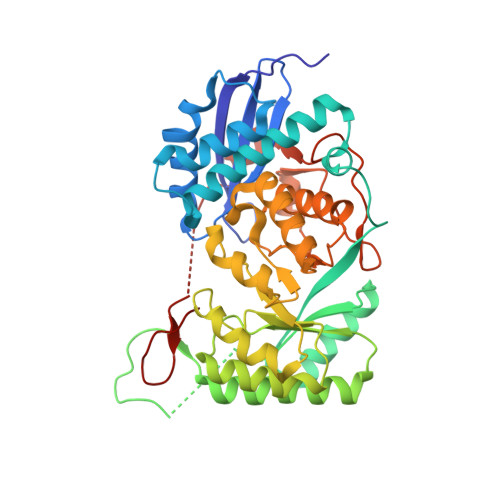 | |
UniProt | |||||
Find proteins for A6M2W4 (Clostridium beijerinckii (strain ATCC 51743 / NCIMB 8052)) Explore A6M2W4 Go to UniProtKB: A6M2W4 | |||||
Entity Groups | |||||
| Sequence Clusters | 30% Identity50% Identity70% Identity90% Identity95% Identity100% Identity | ||||
| UniProt Group | A6M2W4 | ||||
Sequence AnnotationsExpand | |||||
| |||||
| Ligands 1 Unique | |||||
|---|---|---|---|---|---|
| ID | Chains | Name / Formula / InChI Key | 2D Diagram | 3D Interactions | |
| MG Query on MG | C [auth A], D [auth B] | MAGNESIUM ION Mg JLVVSXFLKOJNIY-UHFFFAOYSA-N |  | ||
| Length ( Å ) | Angle ( ˚ ) |
|---|---|
| a = 111.259 | α = 90 |
| b = 111.259 | β = 90 |
| c = 139.803 | γ = 90 |
| Software Name | Purpose |
|---|---|
| ADSC | data collection |
| BALBES | phasing |
| PHENIX | refinement |
| DENZO | data reduction |
| SCALEPACK | data scaling |
RCSB PDB (citation) is hosted by
RCSB PDB is a member of the

