3S3Y

Crystal Structure an Tandem Cyanovirin-N Dimer, CVN2L0


Experimental Data Snapshot

  • Method: X-RAY DIFFRACTION
  • Resolution: 2.00 Å
  • R-Value Free: 0.249 
  • R-Value Work: 0.220 
  • R-Value Observed: 0.221 

wwPDB Validation   3D Report Full Report


This is version 1.4 of the entry. See complete history


Literature

Designed oligomers of cyanovirin-N show enhanced HIV neutralization.

Keeffe, J.R.Gnanapragasam, P.N.Gillespie, S.K.Yong, J.Bjorkman, P.J.Mayo, S.L.

(2011) Proc Natl Acad Sci U S A 108: 14079-14084

  • DOI: https://doi.org/10.1073/pnas.1108777108
  • Primary Citation of Related Structures:  
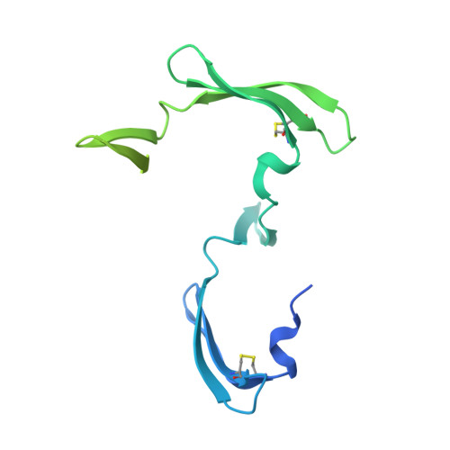
    3S3Y, 3S3Z

  • PubMed Abstract: 

    Cyanovirin-N (CV-N) is a small, cyanobacterial lectin that neutralizes many enveloped viruses, including human immunodeficiency virus type I (HIV-1). This antiviral activity is attributed to two homologous carbohydrate binding sites that specifically bind high mannose glycosylation present on envelope glycoproteins such as HIV-1 gp120. We created obligate CV-N oligomers to determine whether increasing the number of binding sites has an effect on viral neutralization. A tandem repeat of two CV-N molecules (CVN(2)) increased HIV-1 neutralization activity by up to 18-fold compared to wild-type CV-N. In addition, the CVN(2) variants showed extensive cross-clade reactivity and were often more potent than broadly neutralizing anti-HIV antibodies. The improvement in activity and broad cross-strain HIV neutralization exhibited by these molecules holds promise for the future therapeutic utility of these and other engineered CV-N variants.


  • Organizational Affiliation

    Division of Biology, Howard Hughes Medical Institute, Division of Chemistry and Chemical Engineering, California Institute of Technology, Pasadena, CA 91125, USA.


Macromolecules
Find similar proteins by:  (by identity cutoff)  |  3D Structure
Entity ID: 1
MoleculeChains Sequence LengthOrganismDetailsImage
TANDEM CYANOVIRIN-N DIMER CVN2L0213Nostoc ellipsosporumMutation(s): 0 
UniProt
Find proteins for P81180 (Nostoc ellipsosporum)
Explore P81180 
Go to UniProtKB:  P81180
Entity Groups  
Sequence Clusters30% Identity50% Identity70% Identity90% Identity95% Identity100% Identity
UniProt GroupP81180
Sequence Annotations
Expand
  • Reference Sequence
Experimental Data & Validation

Experimental Data

  • Method: X-RAY DIFFRACTION
  • Resolution: 2.00 Å
  • R-Value Free: 0.249 
  • R-Value Work: 0.220 
  • R-Value Observed: 0.221 
  • Space Group: P 32 2 1
Unit Cell:
Length ( Å )Angle ( ˚ )
a = 47.865α = 90
b = 47.865β = 90
c = 78.684γ = 120
Software Package:
Software NamePurpose
MOSFLMdata reduction
SCALAdata scaling
PHASERphasing
REFMACrefinement
PDB_EXTRACTdata extraction
CrystalCleardata collection

Structure Validation

View Full Validation Report


Entry History 

Deposition Data

Revision History  (Full details and data files)

  • Version 1.0: 2011-08-03
    Type: Initial release
  • Version 1.1: 2011-08-10
    Changes: Database references
  • Version 1.2: 2011-09-14
    Changes: Database references
  • Version 1.3: 2017-08-02
    Changes: Advisory, Refinement description, Source and taxonomy
  • Version 1.4: 2023-09-13
    Changes: Advisory, Data collection, Database references, Derived calculations, Refinement description