3S2V

Crystal Structure of the Ligand Binding Domain of GluK1 in Complex with an Antagonist (S)-1-(2'-Amino-2'-carboxyethyl)-3-[(2-carboxythien-3-yl)methyl]thieno[3,4-d]pyrimidin-2,4-dione at 2.5 A Resolution


Experimental Data Snapshot

  • Method: X-RAY DIFFRACTION
  • Resolution: 2.50 Å
  • R-Value Free: 0.257 
  • R-Value Work: 0.178 
  • R-Value Observed: 0.182 

wwPDB Validation   3D Report Full Report


Ligand Structure Quality Assessment 


This is version 1.5 of the entry. See complete history


Literature

Selective kainate receptor (GluK1) ligands structurally based upon 1H-cyclopentapyrimidin-2,4(1H,3H)-dione: synthesis, molecular modeling, and pharmacological and biostructural characterization.

Venskutonyte, R.Butini, S.Coccone, S.S.Gemma, S.Brindisi, M.Kumar, V.Guarino, E.Maramai, S.Valenti, S.Amir, A.Valades, E.A.Frydenvang, K.Kastrup, J.S.Novellino, E.Campiani, G.Pickering, D.S.

(2011) J Med Chem 54: 4793-4805

  • DOI: https://doi.org/10.1021/jm2004078
  • Primary Citation of Related Structures:  
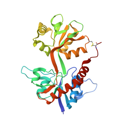
    3S2V

  • PubMed Abstract: 

    The physiological function of kainate receptors (GluK1-GluK5) in the central nervous system is not fully understood yet. With the aim of developing potent and selective GluK1 ligands, we have synthesized a series of new thiophene-based GluK1 agonists (6a-c) and antagonists (7a-d). Pharmacological evaluation revealed that they are selective for the GluK1 subunit, with 7b being the most subtype-selective ligand reported to date (GluK1 vs GluK3). The antagonist 7a was cocrystallized with the GluK1 ligand binding domain, and an X-ray crystallographic analysis revealed the largest flexibility in GluK1 ligand binding domain opening upon binding of a ligand seen to date. The results provide new insights into the molecular mechanism of GluK1 receptor ligand binding and pave the way to the development of new tool compounds for studying kainate receptor function.


  • Organizational Affiliation

    Department of Pharmacology and Pharmacotherapy, Faculty of Pharmaceutical Sciences, University of Copenhagen, Copenhagen, Denmark.


Macromolecules
Find similar proteins by:  (by identity cutoff)  |  3D Structure
Entity ID: 1
MoleculeChains Sequence LengthOrganismDetailsImage
Glutamate receptor, ionotropic kainate 1
A, B
257Rattus norvegicusMutation(s): 0 
Gene Names: GRIK1
UniProt
Find proteins for P22756 (Rattus norvegicus)
Explore P22756 
Go to UniProtKB:  P22756
Entity Groups  
Sequence Clusters30% Identity50% Identity70% Identity90% Identity95% Identity100% Identity
UniProt GroupP22756
Sequence Annotations
Expand
  • Reference Sequence
Small Molecules
Ligands 4 Unique
IDChains Name / Formula / InChI Key2D Diagram3D Interactions
3HU
Query on 3HU

Download Ideal Coordinates CCD File 
C [auth A],
F [auth B]
(S)-1-(2'-AMINO-2'-CARBOXYETHYL)-3-[(2-CARBOXYTHIEN-3-YL)METHYL]THIENO[3,4-D]PYRIMIDIN-2,4-DIONE
C15 H13 N3 O6 S2
LSNSPQPOGKSTHQ-VIFPVBQESA-N
SO4
Query on SO4

Download Ideal Coordinates CCD File 
D [auth A],
G [auth B],
H [auth B]
SULFATE ION
O4 S
QAOWNCQODCNURD-UHFFFAOYSA-L
GOL
Query on GOL

Download Ideal Coordinates CCD File 
E [auth A],
J [auth B]
GLYCEROL
C3 H8 O3
PEDCQBHIVMGVHV-UHFFFAOYSA-N
CL
Query on CL

Download Ideal Coordinates CCD File 
I [auth B]CHLORIDE ION
Cl
VEXZGXHMUGYJMC-UHFFFAOYSA-M
Binding Affinity Annotations 
IDSourceBinding Affinity
3HU BindingDB:  3S2V Ki: min: 157, max: 9.90e+4 (nM) from 2 assay(s)
PDBBind:  3S2V Ki: 157 (nM) from 1 assay(s)
Binding MOAD:  3S2V Ki: 157 (nM) from 1 assay(s)
Experimental Data & Validation

Experimental Data

  • Method: X-RAY DIFFRACTION
  • Resolution: 2.50 Å
  • R-Value Free: 0.257 
  • R-Value Work: 0.178 
  • R-Value Observed: 0.182 
  • Space Group: P 21 21 21
Unit Cell:
Length ( Å )Angle ( ˚ )
a = 61.459α = 90
b = 93.353β = 90
c = 107.171γ = 90
Software Package:
Software NamePurpose
MAR345data collection
PHASERphasing
PHENIXrefinement
XDSdata reduction
SCALAdata scaling

Structure Validation

View Full Validation Report


Ligand Structure Quality Assessment 


Entry History 

Deposition Data

Revision History  (Full details and data files)

  • Version 1.0: 2011-06-22
    Type: Initial release
  • Version 1.1: 2011-07-13
    Changes: Version format compliance
  • Version 1.2: 2012-05-23
    Changes: Database references
  • Version 1.3: 2017-08-09
    Changes: Data collection, Refinement description, Source and taxonomy
  • Version 1.4: 2017-11-08
    Changes: Refinement description
  • Version 1.5: 2023-09-13
    Changes: Data collection, Database references, Derived calculations, Refinement description