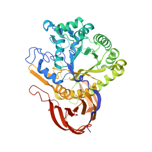
Structure of a novel thermostable GH51 Alpha-L-arabinofuranosidase from Thermotoga petrophila RKU-1
Souza, T.A., Santos, C.R., Souza, A.R., Oldiges, D.P., Ruller, R., Prade, R.A., Squina, F.M., Murakami, M.T.(2011) Protein Sci 20: 1632-1637
- PubMed: 21796714
- DOI: https://doi.org/10.1002/pro.693
- Primary Citation of Related Structures:
3S2C - PubMed Abstract:
α-L-arabinofuranosidases (EC 3.2.1.55) participate in the degradation of a variety of L-arabinose-containing polysaccharides and interact synergistically with other hemicellulases in the production of oligosaccharides and bioconversion of lignocellulosic biomass into biofuels. In this work, the structure of a novel thermostable family 51 (GH51) α-L-arabinofuranosidase from Thermotoga petrophila RKU-1 (TpAraF) was determined at 3.1 Å resolution. The TpAraF tertiary structure consists of an (α/β)-barrel catalytic core associated with a C-terminal β-sandwich domain, which is stabilized by hydrophobic contacts. In contrast to other structurally characterized GH51 AraFs, the accessory domain of TpAraF is intimately linked to the active site by a long β-hairpin motif, which modifies the catalytic cavity in shape and volume. Sequence and structural analyses indicate that this motif is unique to Thermotoga AraFs. Small angle X-ray scattering investigation showed that TpAraF assembles as a hexamer in solution and is preserved at the optimum catalytic temperature, 65°C, suggesting functional significance. Crystal packing analysis shows that the biological hexamer encompasses a dimer of trimers and the multiple oligomeric interfaces are predominantly fashioned by polar and electrostatic contacts.
Organizational Affiliation:
Laboratório Nacional de Biociências, Centro Nacional de Pesquisa em Energia e Materiais, Campinas, São Paulo, Brazil.