3RVA

Crystal structure of organophosphorus acid anhydrolase from Alteromonas macleodii


Experimental Data Snapshot

  • Method: X-RAY DIFFRACTION
  • Resolution: 1.80 Å
  • R-Value Work: 0.156 
  • R-Value Observed: 0.156 

wwPDB Validation   3D Report Full Report


This is version 1.4 of the entry. See complete history


Literature

Organophosphorus acid anhydrolase from Alteromonas macleodii: structural study and functional relationship to prolidases.

Stepankova, A.Duskova, J.Skalova, T.Hasek, J.Koval, T.Ostergaard, L.H.Dohnalek, J.

(2013) Acta Crystallogr Sect F Struct Biol Cryst Commun 69: 346-354

  • DOI: https://doi.org/10.1107/S1744309113002674
  • Primary Citation of Related Structures:  
    3RVA

  • PubMed Abstract: 

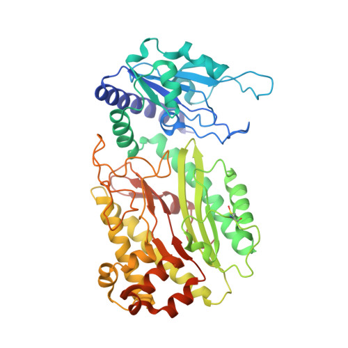
    The bacterial enzyme organophosphorus acid anhydrolase (OPAA) is able to catalyze the hydrolysis of both proline dipeptides (Xaa-Pro) and several types of organophosphate (OP) compounds. The full three-dimensional structure of the manganese-dependent OPAA enzyme is presented for the first time. This enzyme, which was originally isolated from the marine bacterium Alteromonas macleodii, was prepared recombinantly in Escherichia coli. The crystal structure was determined at 1.8 Å resolution in space group C2, with unit-cell parameters a = 133.8, b = 49.2, c = 97.3 Å, β = 125.0°. The enzyme forms dimers and their existence in solution was confirmed by dynamic light scattering and size-exclusion chromatography. The enzyme shares the pita-bread fold of its C-terminal domain with related prolidases. The binuclear manganese centre is located in the active site within the pita-bread domain. Moreover, an Ni(2+) ion from purification was localized according to anomalous signal. This study presents the full structure of this enzyme with complete surroundings of the active site and provides a critical analysis of its relationship to prolidases.


  • Organizational Affiliation

    Institute of Macromolecular Chemistry, AS CR, vvi, Heyrovsky sq 2, 162 06 Prague 6, Czech Republic. a.stepanko@gmail.com


Macromolecules
Find similar proteins by:  (by identity cutoff)  |  3D Structure
Entity ID: 1
MoleculeChains Sequence LengthOrganismDetailsImage
Organophosphorus acid anhydrolase451Alteromonas macleodiiMutation(s): 0 
EC: 3.1.8.2 (PDB Primary Data), 3.4.13.9 (PDB Primary Data)
UniProt
Find proteins for F8UVJ4 (Alteromonas macleodii)
Explore F8UVJ4 
Go to UniProtKB:  F8UVJ4
Entity Groups  
Sequence Clusters30% Identity50% Identity70% Identity90% Identity95% Identity100% Identity
UniProt GroupF8UVJ4
Sequence Annotations
Expand
  • Reference Sequence
Experimental Data & Validation

Experimental Data

  • Method: X-RAY DIFFRACTION
  • Resolution: 1.80 Å
  • R-Value Work: 0.156 
  • R-Value Observed: 0.156 
  • Space Group: C 1 2 1
Unit Cell:
Length ( Å )Angle ( ˚ )
a = 133.77α = 90
b = 49.19β = 125.03
c = 97.34γ = 90
Software Package:
Software NamePurpose
MxCuBEdata collection
MOLREPphasing
REFMACrefinement
HKL-2000data reduction
HKL-2000data scaling

Structure Validation

View Full Validation Report


Entry History 

Deposition Data

Revision History  (Full details and data files)

  • Version 1.0: 2012-05-09
    Type: Initial release
  • Version 1.1: 2013-04-10
    Changes: Database references
  • Version 1.2: 2018-01-31
    Changes: Database references
  • Version 1.3: 2023-09-13
    Changes: Data collection, Database references, Derived calculations, Refinement description
  • Version 1.4: 2023-12-06
    Changes: Data collection