3RJD

Crystal structure of Fc RI and its implication to high affinity immunoglobulin G binding


Experimental Data Snapshot

  • Method: X-RAY DIFFRACTION
  • Resolution: 2.65 Å
  • R-Value Free: 0.276 
  • R-Value Work: 0.248 
  • R-Value Observed: 0.250 

wwPDB Validation   3D Report Full Report


Ligand Structure Quality Assessment 


This is version 2.0 of the entry. See complete history


Literature

Crystal structure of Fc gamma receptor I and its implication in high affinity gamma-immunoglobulin binding.

Lu, J.Ellsworth, J.L.Hamacher, N.Oak, S.W.Sun, P.D.

(2011) J Biol Chem 286: 40608-40613

  • DOI: https://doi.org/10.1074/jbc.M111.257550
  • Primary Citation of Related Structures:  
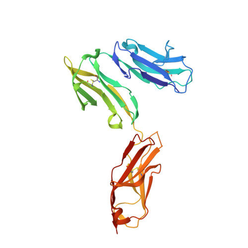
    3RJD

  • PubMed Abstract: 

    Fcγ receptors (FcγRs) play critical roles in humoral and cellular immune responses through interactions with the Fc region of immunoglobulin G (IgG). Among them, FcγRI is the only high affinity receptor for IgG and thus is a potential target for immunotherapy. Here we report the first crystal structure of an FcγRI with all three extracellular Ig-like domains (designated as D1, D2, and D3). The structure shows that, first, FcγRI has an acute D1-D2 hinge angle similar to that of FcεRI but much smaller than those observed in the low affinity Fcγ receptors. Second, the D3 domain of FcγRI is positioned away from the putative IgG binding site on the receptor and is thus unlikely to make direct contacts with Fc. Third, the replacement of FcγRIII FG-loop ((171)LVGSKNV(177)) with that of FcγRI ((171)MGKHRY(176)) resulted in a 15-fold increase in IgG(1) binding affinity, whereas a valine insertion in the FcγRI FG-loop ((171)MVGKHRY(177)) abolished the affinity enhancement. Thus, the FcγRI FG-loop with its conserved one-residue deletion is critical to the high affinity IgG binding. The structural results support FcγRI binding to IgG in a similar mode as its low affinity counterparts. Taken together, our study suggests a molecular mechanism for the high affinity IgG recognition by FcγRI and provides a structural basis for understanding its physiological function and its therapeutic implication in treating autoimmune diseases.


  • Organizational Affiliation

    Structural Immunology Section, Laboratory of Immunogenetics, NIAID, National Institutes of Health, Rockville, Maryland, USA.


Macromolecules
Find similar proteins by:  (by identity cutoff)  |  3D Structure
Entity ID: 1
MoleculeChains Sequence LengthOrganismDetailsImage
High affinity immunoglobulin gamma Fc receptor I262Homo sapiensMutation(s): 0 
Gene Names: FCGR1AFCG1FCGR1IGFR1
UniProt & NIH Common Fund Data Resources
Find proteins for P12314 (Homo sapiens)
Explore P12314 
Go to UniProtKB:  P12314
PHAROS:  P12314
GTEx:  ENSG00000150337 
Entity Groups  
Sequence Clusters30% Identity50% Identity70% Identity90% Identity95% Identity100% Identity
UniProt GroupP12314
Sequence Annotations
Expand
  • Reference Sequence
Oligosaccharides

Help

Entity ID: 2
MoleculeChains Length2D Diagram Glycosylation3D Interactions
alpha-D-mannopyranose-(1-4)-2-acetamido-2-deoxy-beta-D-glucopyranose-(1-4)-2-acetamido-2-deoxy-beta-D-glucopyranose
B
3N-Glycosylation
Glycosylation Resources
GlyTouCan:  G62182OO
GlyCosmos:  G62182OO
GlyGen:  G62182OO
Entity ID: 3
MoleculeChains Length2D Diagram Glycosylation3D Interactions
alpha-D-mannopyranose-(1-4)-2-acetamido-2-deoxy-beta-D-glucopyranose-(1-4)-[alpha-L-fucopyranose-(1-6)]2-acetamido-2-deoxy-beta-D-glucopyranose
C
4N-Glycosylation
Glycosylation Resources
GlyTouCan:  G47477HI
GlyCosmos:  G47477HI
GlyGen:  G47477HI
Entity ID: 4
MoleculeChains Length2D Diagram Glycosylation3D Interactions
alpha-D-mannopyranose-(1-4)-2-acetamido-2-deoxy-beta-D-glucopyranose
D
2N-Glycosylation
Glycosylation Resources
GlyTouCan:  G35893LO
GlyCosmos:  G35893LO
GlyGen:  G35893LO
Entity ID: 5
MoleculeChains Length2D Diagram Glycosylation3D Interactions
2-acetamido-2-deoxy-beta-D-glucopyranose-(1-4)-2-acetamido-2-deoxy-beta-D-glucopyranose
E
2N-Glycosylation
Glycosylation Resources
GlyTouCan:  G42666HT
GlyCosmos:  G42666HT
GlyGen:  G42666HT
Small Molecules
Experimental Data & Validation

Experimental Data

  • Method: X-RAY DIFFRACTION
  • Resolution: 2.65 Å
  • R-Value Free: 0.276 
  • R-Value Work: 0.248 
  • R-Value Observed: 0.250 
  • Space Group: P 32 2 1
Unit Cell:
Length ( Å )Angle ( ˚ )
a = 92.828α = 90
b = 92.828β = 90
c = 90.763γ = 120
Software Package:
Software NamePurpose
HKL-2000data collection
BALBESphasing
PHASERphasing
BUSTERrefinement
HKL-2000data reduction
HKL-2000data scaling

Structure Validation

View Full Validation Report


Ligand Structure Quality Assessment 


Entry History 

Deposition Data

Revision History  (Full details and data files)

  • Version 1.0: 2011-09-21
    Type: Initial release
  • Version 1.1: 2013-08-28
    Changes: Database references
  • Version 1.2: 2017-11-08
    Changes: Refinement description
  • Version 2.0: 2020-07-29
    Type: Remediation
    Reason: Carbohydrate remediation
    Changes: Advisory, Atomic model, Data collection, Derived calculations, Structure summary