3RDU

Crystal structure of R7-2 streptavidin complexed with PEG


Experimental Data Snapshot

  • Method: X-RAY DIFFRACTION
  • Resolution: 1.50 Å
  • R-Value Free: 0.207 
  • R-Value Work: 0.170 
  • R-Value Observed: 0.171 

wwPDB Validation   3D Report Full Report


This is version 1.3 of the entry. See complete history


Literature

Evolved streptavidin mutants reveal key role of loop residue in high-affinity binding.

Magalhaes, M.L.Czekster, C.M.Guan, R.Malashkevich, V.N.Almo, S.C.Levy, M.

(2011) Protein Sci 20: 1145-1154

  • DOI: https://doi.org/10.1002/pro.642
  • Primary Citation of Related Structures:  
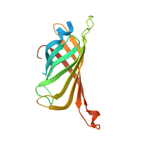
    3RDM, 3RDO, 3RDQ, 3RDS, 3RDU, 3RDX, 3RE5, 3RE6

  • PubMed Abstract: 

    We have performed a detailed analysis of streptavidin variants with altered specificity towards desthiobiotin. In addition to changes in key residues which widen the ligand binding pocket and accommodate the more structurally flexible desthiobiotin, the data revealed the role of a key, non-active site mutation at the base of the flexible loop (S52G) which slows dissociation of this ligand by approximately sevenfold. Our data suggest that this mutation results in the loss of a stabilizing contact which keeps this loop open and accessible in the absence of ligand. When this mutation was introduced into the wild-type protein, destabilization of the opened loop conferred a ∼10-fold decrease in both the on-rate and off-rate for the ligand biotin-4-fluoroscein. A similar effect was observed when this mutation was added to a monomeric form of this protein. Our results provide key insight into the role of the streptavidin flexible loop in ligand binding and maintaining high affinity interactions.


  • Organizational Affiliation

    Department of Biochemistry, Albert Einstein College of Medicine, Bronx, New York 10461, USA.


Macromolecules
Find similar proteins by:  (by identity cutoff)  |  3D Structure
Entity ID: 1
MoleculeChains Sequence LengthOrganismDetailsImage
Streptavidin153Streptomyces avidiniiMutation(s): 6 
UniProt
Find proteins for P22629 (Streptomyces avidinii)
Explore P22629 
Go to UniProtKB:  P22629
Entity Groups  
Sequence Clusters30% Identity50% Identity70% Identity90% Identity95% Identity100% Identity
UniProt GroupP22629
Sequence Annotations
Expand
  • Reference Sequence
Experimental Data & Validation

Experimental Data

  • Method: X-RAY DIFFRACTION
  • Resolution: 1.50 Å
  • R-Value Free: 0.207 
  • R-Value Work: 0.170 
  • R-Value Observed: 0.171 
  • Space Group: P 42 21 2
Unit Cell:
Length ( Å )Angle ( ˚ )
a = 57.354α = 90
b = 57.354β = 90
c = 76.839γ = 90
Software Package:
Software NamePurpose
SCALEPACKdata scaling
PHASERphasing
REFMACrefinement
PDB_EXTRACTdata extraction
CBASSdata collection
HKL-2000data reduction

Structure Validation

View Full Validation Report


Entry History 

Deposition Data

Revision History  (Full details and data files)

  • Version 1.0: 2011-07-06
    Type: Initial release
  • Version 1.1: 2011-07-13
    Changes: Version format compliance
  • Version 1.2: 2018-01-24
    Changes: Structure summary
  • Version 1.3: 2023-09-13
    Changes: Data collection, Database references, Derived calculations, Refinement description