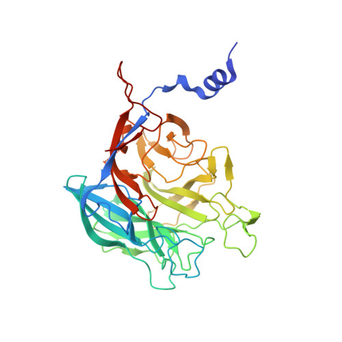
3R4Y

Crystal structure of alpha-neoagarobiose hydrolase (ALPHA-NABH) from Saccharophagus degradans 2-40


Experimental Data Snapshot

  • Method: X-RAY DIFFRACTION
  • Resolution: 2.00 Å
  • R-Value Free: 0.206 
  • R-Value Work: 0.161 
  • R-Value Observed: 0.164 

wwPDB Validation   3D Report Full Report


This is version 1.1 of the entry. See complete history


Literature

Crystal structure of a key enzyme in the agarolytic pathway, alpha-neoagarobiose hydrolase from Saccharophagus degradans 2-40

Ha, S.C.Lee, S.Lee, J.Kim, H.T.Ko, H.J.Kim, K.H.Choi, I.G.

(2011) Biochem Biophys Res Commun 412: 238-244

  • DOI: https://doi.org/10.1016/j.bbrc.2011.07.073
  • Primary Citation of Related Structures:  
    3R4Y

  • PubMed Abstract: 

    In agarolytic microorganisms, α-neoagarobiose hydrolase (NABH) is an essential enzyme to metabolize agar because it converts α-neoagarobiose (O-3,6-anhydro-alpha-l-galactopyranosyl-(1,3)-d-galactose) into fermentable monosaccharides (d-galactose and 3,6-anhydro-l-galactose) in the agarolytic pathway. NABH can be divided into two biological classes by its cellular location. Here, we describe a structure and function of cytosolic NABH from Saccharophagus degradans 2-40 in a native protein and d-galactose complex determined at 2.0 and 1.55 Å, respectively. The overall fold is organized in an N-terminal helical extension and a C-terminal five-bladed β-propeller catalytic domain. The structure of the enzyme-ligand (d-galactose) complex predicts a +1 subsite in the substrate binding pocket. The structural features may provide insights for the evolution and classification of NABH in agarolytic pathways.


  • Organizational Affiliation

    Physical Bioscience Division, Lawrence Berkeley National Laboratory, Berkeley, CA 94720, USA.


Macromolecules
Find similar proteins by:  (by identity cutoff)  |  3D Structure
Entity ID: 1
MoleculeChains Sequence LengthOrganismDetailsImage
Glycosyl hydrolase family 32, N terminal
A, B
374Saccharophagus degradans 2-40Mutation(s): 0 
Gene Names: Sde_2657
EC: 3.2.1
UniProt
Find proteins for Q21HB2 (Saccharophagus degradans (strain 2-40 / ATCC 43961 / DSM 17024))
Explore Q21HB2 
Go to UniProtKB:  Q21HB2
Entity Groups  
Sequence Clusters30% Identity50% Identity70% Identity90% Identity95% Identity100% Identity
UniProt GroupQ21HB2
Sequence Annotations
Expand
  • Reference Sequence
Experimental Data & Validation

Experimental Data

  • Method: X-RAY DIFFRACTION
  • Resolution: 2.00 Å
  • R-Value Free: 0.206 
  • R-Value Work: 0.161 
  • R-Value Observed: 0.164 
  • Space Group: C 1 2 1
Unit Cell:
Length ( Å )Angle ( ˚ )
a = 129.87α = 90
b = 76.81β = 101.86
c = 90.11γ = 90
Software Package:
Software NamePurpose
ADSCdata collection
SOLVEphasing
REFMACrefinement
HKL-2000data reduction
HKL-2000data scaling

Structure Validation

View Full Validation Report


Entry History 

Deposition Data

Revision History  (Full details and data files)

  • Version 1.0: 2012-02-01
    Type: Initial release
  • Version 1.1: 2024-03-20
    Changes: Data collection, Database references