3R2P

2.2 Angstrom Crystal Structure of C Terminal Truncated Human Apolipoprotein A-I Reveals the Assembly of HDL by Dimerization.


Experimental Data Snapshot

  • Method: X-RAY DIFFRACTION
  • Resolution: 2.20 Å
  • R-Value Free: 0.280 
  • R-Value Work: 0.230 
  • R-Value Observed: 0.236 

wwPDB Validation   3D Report Full Report


This is version 1.4 of the entry. See complete history


Literature

Crystal Structure of C-terminal Truncated Apolipoprotein A-I Reveals the Assembly of High Density Lipoprotein (HDL) by Dimerization.

Mei, X.Atkinson, D.

(2011) J Biol Chem 286: 38570-38582

  • DOI: https://doi.org/10.1074/jbc.M111.260422
  • Primary Citation of Related Structures:  
    3R2P

  • PubMed Abstract: 

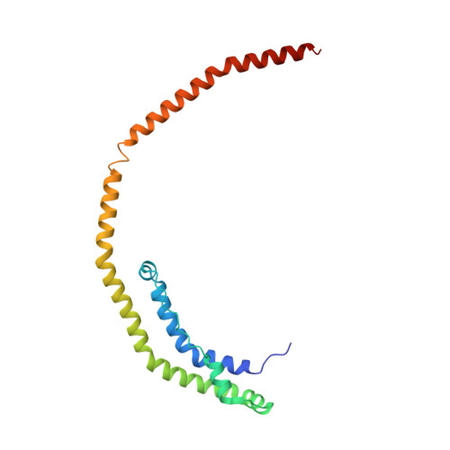
    Apolipoprotein A-I (apoA-I) plays important structural and functional roles in plasma high density lipoprotein (HDL) that is responsible for reverse cholesterol transport. However, a molecular understanding of HDL assembly and function remains enigmatic. The 2.2-Å crystal structure of Δ(185-243)apoA-I reported here shows that it forms a half-circle dimer. The backbone of the dimer consists of two elongated antiparallel proline-kinked helices (five AB tandem repeats). The N-terminal domain of each molecule forms a four-helix bundle with the helical C-terminal region of the symmetry-related partner. The central region forms a flexible domain with two antiparallel helices connecting the bundles at each end. The two-domain dimer structure based on helical repeats suggests the role of apoA-I in the formation of discoidal HDL particles. Furthermore, the structure suggests the possible interaction with lecithin-cholesterol acyltransferase and may shed light on the molecular details of the effect of the Milano, Paris, and Fin mutations.


  • Organizational Affiliation

    Department of Physiology and Biophysics, Boston University School of Medicine, Boston, Massachusetts 02118.


Macromolecules
Find similar proteins by:  (by identity cutoff)  |  3D Structure
Entity ID: 1
MoleculeChains Sequence LengthOrganismDetailsImage
Apolipoprotein A-I185Homo sapiensMutation(s): 0 
Gene Names: APOA1
UniProt & NIH Common Fund Data Resources
Find proteins for P02647 (Homo sapiens)
Explore P02647 
Go to UniProtKB:  P02647
PHAROS:  P02647
GTEx:  ENSG00000118137 
Entity Groups  
Sequence Clusters30% Identity50% Identity70% Identity90% Identity95% Identity100% Identity
UniProt GroupP02647
Sequence Annotations
Expand
  • Reference Sequence
Experimental Data & Validation

Experimental Data

  • Method: X-RAY DIFFRACTION
  • Resolution: 2.20 Å
  • R-Value Free: 0.280 
  • R-Value Work: 0.230 
  • R-Value Observed: 0.236 
  • Space Group: P 21 21 2
Unit Cell:
Length ( Å )Angle ( ˚ )
a = 108.333α = 90
b = 30.178β = 90
c = 69.576γ = 90
Software Package:
Software NamePurpose
MAR345dtbdata collection
PHENIXmodel building
PHENIXrefinement
HKL-2000data reduction
HKL-2000data scaling
PHENIXphasing

Structure Validation

View Full Validation Report


Entry History 

Deposition Data

Revision History  (Full details and data files)

  • Version 1.0: 2011-09-21
    Type: Initial release
  • Version 1.1: 2011-09-28
    Changes: Database references, Structure summary
  • Version 1.2: 2011-11-16
    Changes: Database references
  • Version 1.3: 2024-02-21
    Changes: Data collection, Database references
  • Version 1.4: 2024-04-03
    Changes: Refinement description