Crystal structures and putative interface of Saccharomyces cerevisiae mitochondrial matrix proteins Mmf1 and Mam33.
Pu, Y.G., Jiang, Y.L., Ye, X.D., Ma, X.X., Guo, P.C., Lian, F.M., Teng, Y.B., Chen, Y., Zhou, C.Z.(2011) J Struct Biol 175: 469-474
- PubMed: 21600990
- DOI: https://doi.org/10.1016/j.jsb.2011.05.005
- Primary Citation of Related Structures:
3QUW, 3QV0 - PubMed Abstract:
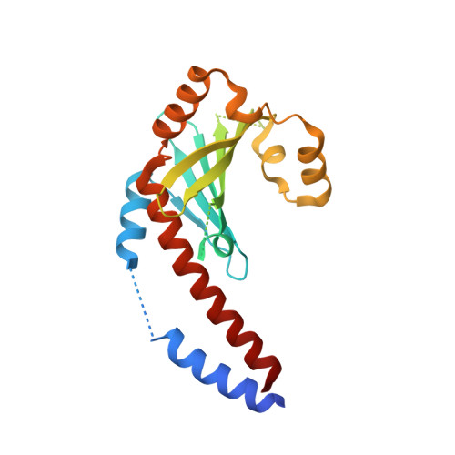
The yeast Saccharomyces cerevisiae mitochondrial matrix factor Mmf1, a member in the YER057c/Yigf/Uk114 family, participates in isoleucine biosynthesis and mitochondria maintenance. Mmf1 physically interacts with another mitochondrial matrix protein Mam33, which is involved in the sorting of cytochrome b₂ to the intermembrane space as well as mitochondrial ribosomal protein synthesis. To elucidate the structural basis for their interaction, we determined the crystal structures of Mmf1 and Mam33 at 1.74 and 2.10 Å, respectively. Both Mmf1 and Mam33 adopt a trimeric structure: each subunit of Mmf1 displays a chorismate mutase fold with a six-stranded β-sheet flanked by two α-helices on one side, whereas a subunit of Mam33 consists of a twisted six-stranded β-sheet surrounded by five α-helices. Biochemical assays combined with structure-based computational simulation enable us to model a putative complex of Mmf1-Mam33, which consists of one Mam33 trimer and two tandem Mmf1 trimers in a head-to-tail manner. The two interfaces between the ring-like trimers are mainly composed of electrostatic interactions mediated by complementary negatively and positively charged patches. These results provided the structural insights into the putative function of Mmf1 during mitochondrial protein synthesis via Mam33, a protein binding to mitochondrial ribosomal proteins.
Organizational Affiliation:
Hefei National Laboratory for Physical Sciences at Microscale and School of Life Sciences, People's Republic of China.