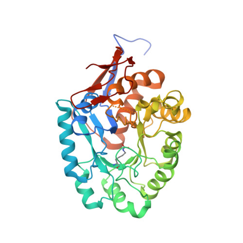
2.4 Angstrom Crystal Structure of Dihydroorotase (pyrC) from Campylobacter jejuni.
Minasov, G., Halavaty, A., Shuvalova, L., Dubrovska, I., Winsor, J., Papazisi, L., Anderson, W.F., Center for Structural Genomics of Infectious Diseases (CSGID)To be published.