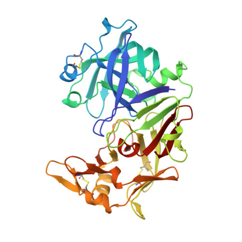
Revised 2.3 A structure of porcine pepsin: evidence for a flexible subdomain
Abad-Zapatero, C., Rydel, T.J., Erickson, J.(1990) Proteins 8: 62-81
- PubMed: 2217165
- DOI: https://doi.org/10.1002/prot.340080109
- Primary Citation of Related Structures:
3PEP - PubMed Abstract:
A revised three-dimensional crystal structure of ethanol-inhibited porcine pepsin refined to an R-factor of 0.171 at 2.3 A resolution is presented and compared to the refined structures of the fungal aspartic proteinases: penicillopepsin, rhizopuspepsin, and endothiapepsin. Pepsin is composed of two nearly equal N and C domains related by an intra dyad. The overall polypeptide fold and active site structures are homologous for pepsin and the fungal enzymes. The weak inhibition of pepsin by ethanol can be explained by the presence of one or more ethanol molecules, in the vicinity of the active site carboxylates, which slightly alter the hydrogen-bonding network and which may compete with substrate binding in the active site. Structural superposition analysis showed that the N domains aligned better than the C-domains for pepsin and the fungal aspartic proteinases: 107-140 C alpha pairs aligned to 0.72-0.85 A rms for the N domains; 64-95 C alpha pairs aligned to 0.78-1.03 A rms for the C domains. The major structural difference between pepsin and the fungal enzymes concerns a newly described subdomain whose conformation varies markedly among these enzyme structures. The subdomain in pepsin comprises nearly 100 residues and is composed of two contiguous segments within the C domain (residues 192-212 and 223-299). the subdomain is connected, or "hinged," to a mixed beta-sheet that forms one of the structurally invariant, active site psi-loops. Relative subdomain displacements as large as a 21.0 degrees rotation and a 5.9 A translation were observed among the different enzymes. There is some suggestion in pepsin that the subdomain may be flexible and perhaps plays a structural role in mediating substrate binding, determining the substrate specificity, or in the activation of the zymogen.
Organizational Affiliation:
Protein Crystallography Laboratory, Abbott Laboratories, Abbott Park, Illinois 60064.