The Conformational Flexibility of the Helicase-like Domain from Thermotoga maritima Reverse Gyrase Is Restricted by the Topoisomerase Domain.
Del Toro Duany, Y., Klostermeier, D., Rudolph, M.G.(2011) Biochemistry 50: 5816-5823
- PubMed: 21627332
- DOI: https://doi.org/10.1021/bi200236a
- Primary Citation of Related Structures:
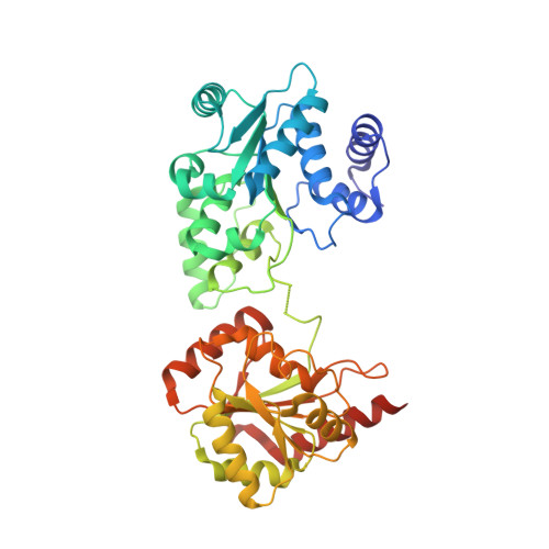
3P4X, 3P4Y - PubMed Abstract:
Reverse gyrase is the only enzyme known to introduce positive supercoils into DNA. Positive supercoiling is achieved by the functional cooperation of a helicase-like and a topoisomerase domain. The isolated helicase-like domain is a DNA-stimulated ATPase, and the isolated topoisomerase domain can relax supercoiled DNA. In the context of reverse gyrase, these individual activities are suppressed or attenuated. The helicase-like domain of Thermotoga maritima reverse gyrase is a nucleotide-dependent conformational switch that binds DNA and ATP cooperatively. It provides a nucleotide-dependent DNA-binding site to reverse gyrase and thus serves as a valuable model for the investigation of the effect of nucleotides on DNA processing by reverse gyrase that is key to its supercoiling activity. To improve our understanding of the structural basis for the functional cooperation of a helicase domain with a DNA topoisomerase, we have determined the structures of the isolated helicase-like domain of T. maritima reverse gyrase in five different conformations. Comparison of these structures reveals extensive domain flexibility in the absence of conformational restrictions by the topoisomerase that is consistent with single-molecule Förster resonance energy transfer experiments presented here. The structure of the first ADP-bound form provides novel details about nucleotide binding to reverse gyrase. It demonstrates that reverse gyrases use the canonical nucleotide binding mode common to superfamily 2 helicases despite large deviations in the conserved motifs. A characteristic insert region adopts drastically different structures in different reverse gyrases. Counterparts of this insert region are located at very different positions in other DNA-processing enzymes but may point toward a general role in DNA strand separation.
Organizational Affiliation:
Biozentrum, Department of Biophysical Chemistry, University of Basel, Klingelbergstrasse 70, CH-4056 Basel, Switzerland.