3OSV

The crytsal structure of FLGD from P. Aeruginosa


Experimental Data Snapshot

  • Method: X-RAY DIFFRACTION
  • Resolution: 2.35 Å
  • R-Value Free: 0.268 
  • R-Value Work: 0.211 
  • R-Value Observed: 0.214 

wwPDB Validation   3D Report Full Report


This is version 1.3 of the entry. See complete history


Literature

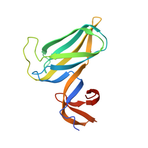
Crystal structure of a novel dimer form of FlgD from P. aeruginosa PAO1

Zhou, H.Luo, M.Cai, X.Tang, J.Niu, S.Zhang, W.Hu, Y.Yin, Y.Huang, A.Wang, D.Wang, D.

(2011) Proteins 79: 2346-2351


Macromolecules
Find similar proteins by:  (by identity cutoff)  |  3D Structure
Entity ID: 1
MoleculeChains Sequence LengthOrganismDetailsImage
Flagellar basal-body rod modification protein FlgD
A, B, C, D
138Pseudomonas aeruginosaMutation(s): 0 
Gene Names: flgDPA1079
UniProt
Find proteins for Q9I4Q0 (Pseudomonas aeruginosa (strain ATCC 15692 / DSM 22644 / CIP 104116 / JCM 14847 / LMG 12228 / 1C / PRS 101 / PAO1))
Explore Q9I4Q0 
Go to UniProtKB:  Q9I4Q0
Entity Groups  
Sequence Clusters30% Identity50% Identity70% Identity90% Identity95% Identity100% Identity
UniProt GroupQ9I4Q0
Sequence Annotations
Expand
  • Reference Sequence
Experimental Data & Validation

Experimental Data

  • Method: X-RAY DIFFRACTION
  • Resolution: 2.35 Å
  • R-Value Free: 0.268 
  • R-Value Work: 0.211 
  • R-Value Observed: 0.214 
  • Space Group: I 2 2 2
Unit Cell:
Length ( Å )Angle ( ˚ )
a = 114.828α = 90
b = 118.379β = 90
c = 118.301γ = 90
Software Package:
Software NamePurpose
SOLVEphasing
REFMACrefinement
HKL-2000data reduction
HKL-2000data scaling

Structure Validation

View Full Validation Report


Entry History 

Deposition Data

Revision History  (Full details and data files)

  • Version 1.0: 2011-05-04
    Type: Initial release
  • Version 1.1: 2011-07-13
    Changes: Version format compliance
  • Version 1.2: 2014-03-26
    Changes: Database references
  • Version 1.3: 2024-03-20
    Changes: Data collection, Database references, Derived calculations