3OPY

Crystal structure of Pichia pastoris phosphofructokinase in the T-state


Experimental Data Snapshot

  • Method: X-RAY DIFFRACTION
  • Resolution: 3.05 Å
  • R-Value Free: 0.232 
  • R-Value Work: 0.200 
  • R-Value Observed: 0.202 

wwPDB Validation   3D Report Full Report


Ligand Structure Quality Assessment 


This is version 1.2 of the entry. See complete history


Literature

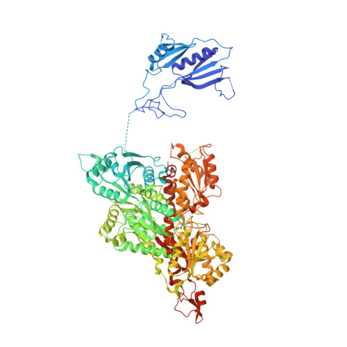
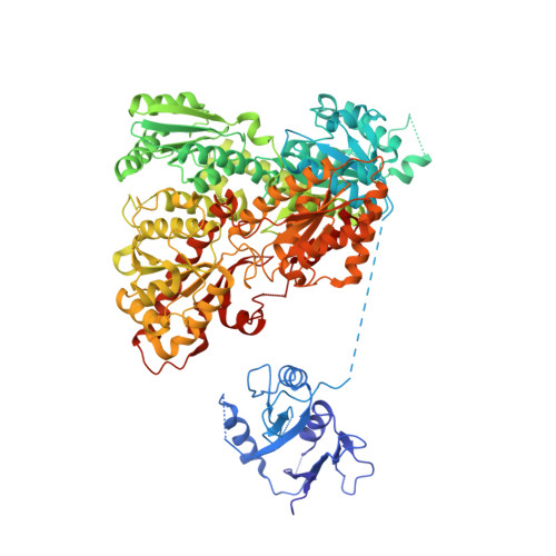
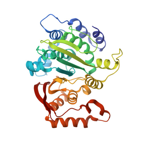
Molecular architecture and structural basis of allosteric regulation of eukaryotic phosphofructokinases.

Strater, N.Marek, S.Kuettner, E.B.Kloos, M.Keim, A.Bruser, A.Kirchberger, J.Schoneberg, T.

(2011) FASEB J 25: 89-98

  • DOI: https://doi.org/10.1096/fj.10-163865
  • Primary Citation of Related Structures:  
    3OPY

  • PubMed Abstract: 

    Eukaryotic ATP-dependent 6-phosphofructokinases (Pfks) differ from their bacterial counterparts in a much more complex structural organization and allosteric regulation. Pichia pastoris Pfk (PpPfk) is, with ∼ 1 MDa, the most complex and probably largest eukaryotic Pfk. We have determined the crystal structure of full-length PpPfk to 3.05 Å resolution in the T state. PpPfk forms a (αβγ)(4) dodecamer of D(2) symmetry with dimensions of 161 × 157 × 233 Å mainly via interactions of the α chains. The N-terminal domains of the α and β chains have folds that are distantly related to glyoxalase I, but the active sites are no longer functional. Interestingly, these domains located at the 2 distal ends of this protein along the long 2-fold axis form a (αβ)(2) dimer as does the core Pfk domains; however, the domains are swapped across the tetramerization interface. In PpPfk, the unique γ subunit participates in oligomerization of the αβ chains. This modulator protein was acquired from an ancient S-adenosylmethionine-dependent methyltransferase. The identification of novel ATP binding sites, which do not correspond to the bacterial catalytic or effector binding sites, point to marked structural and functional differences between bacterial and eukaryotic Pfks.


  • Organizational Affiliation

    Institute of Bioanalytical Chemistry, Center for Biotechnology and Biomedicine, University of Leipzig, Deutscher Platz 5, 04103 Leipzig, Germany. strater@bbz.uni-leipzig.de


Macromolecules
Find similar proteins by:  (by identity cutoff)  |  3D Structure
Entity ID: 1
MoleculeChains Sequence LengthOrganismDetailsImage
6-phosphofructo-1-kinase alpha-subunit
A, C, E, G
989Komagataella pastoris DSMZ 70382Mutation(s): 0 
EC: 2.7.1.11
UniProt
Find proteins for Q8NJU8 (Komagataella pastoris)
Explore Q8NJU8 
Go to UniProtKB:  Q8NJU8
Entity Groups  
Sequence Clusters30% Identity50% Identity70% Identity90% Identity95% Identity100% Identity
UniProt GroupQ8NJU8
Sequence Annotations
Expand
  • Reference Sequence
Find similar proteins by:  (by identity cutoff)  |  3D Structure
Entity ID: 2
MoleculeChains Sequence LengthOrganismDetailsImage
6-phosphofructo-1-kinase beta-subunit
B, D, F, H
941Komagataella pastoris DSMZ 70382Mutation(s): 0 
EC: 2.7.1.11
UniProt
Find proteins for Q8TGA0 (Komagataella pastoris)
Explore Q8TGA0 
Go to UniProtKB:  Q8TGA0
Entity Groups  
Sequence Clusters30% Identity50% Identity70% Identity90% Identity95% Identity100% Identity
UniProt GroupQ8TGA0
Sequence Annotations
Expand
  • Reference Sequence
Find similar proteins by:  (by identity cutoff)  |  3D Structure
Entity ID: 3
MoleculeChains Sequence LengthOrganismDetailsImage
6-phosphofructo-1-kinase gamma-subunit
I, J, K, L
351Komagataella pastoris DSMZ 70382Mutation(s): 0 
EC: 2.7.1.11
UniProt
Find proteins for A7MAS3 (Komagataella pastoris)
Explore A7MAS3 
Go to UniProtKB:  A7MAS3
Entity Groups  
Sequence Clusters30% Identity50% Identity70% Identity90% Identity95% Identity100% Identity
UniProt GroupA7MAS3
Sequence Annotations
Expand
  • Reference Sequence
Small Molecules
Ligands 2 Unique
IDChains Name / Formula / InChI Key2D Diagram3D Interactions
ATP
Query on ATP

Download Ideal Coordinates CCD File 
KB [auth H],
MA [auth D],
YA [auth F],
Z [auth B]
ADENOSINE-5'-TRIPHOSPHATE
C10 H16 N5 O13 P3
ZKHQWZAMYRWXGA-KQYNXXCUSA-N
SO4
Query on SO4

Download Ideal Coordinates CCD File 
AA [auth B]
AB [auth F]
AC [auth L]
BA [auth B]
BB [auth F]
AA [auth B],
AB [auth F],
AC [auth L],
BA [auth B],
BB [auth F],
BC [auth L],
CA [auth C],
CB [auth G],
CC [auth L],
DA [auth C],
DB [auth G],
EA [auth C],
EB [auth G],
FA [auth C],
FB [auth G],
GA [auth C],
GB [auth G],
HA [auth C],
HB [auth G],
IA [auth C],
IB [auth G],
JA [auth C],
JB [auth G],
KA [auth C],
LA [auth C],
LB [auth H],
M [auth A],
MB [auth H],
N [auth A],
NA [auth D],
NB [auth H],
O [auth A],
OA [auth D],
OB [auth H],
P [auth A],
PA [auth D],
PB [auth I],
Q [auth A],
QA [auth E],
QB [auth I],
R [auth A],
RA [auth E],
RB [auth I],
S [auth A],
SA [auth E],
SB [auth I],
T [auth A],
TA [auth E],
TB [auth I],
U [auth A],
UA [auth E],
UB [auth J],
V [auth A],
VA [auth E],
VB [auth J],
W [auth A],
WA [auth E],
WB [auth J],
X [auth A],
XA [auth E],
XB [auth K],
Y [auth A],
YB [auth K],
ZA [auth F],
ZB [auth K]
SULFATE ION
O4 S
QAOWNCQODCNURD-UHFFFAOYSA-L
Experimental Data & Validation

Experimental Data

  • Method: X-RAY DIFFRACTION
  • Resolution: 3.05 Å
  • R-Value Free: 0.232 
  • R-Value Work: 0.200 
  • R-Value Observed: 0.202 
  • Space Group: P 1 21 1
Unit Cell:
Length ( Å )Angle ( ˚ )
a = 161.658α = 90
b = 188.303β = 92.85
c = 231.56γ = 90
Software Package:
Software NamePurpose
MAR345dtbdata collection
MOLREPphasing
BUSTERrefinement
XDSdata reduction
XDSdata scaling

Structure Validation

View Full Validation Report


Ligand Structure Quality Assessment 


Entry History 

Deposition Data

Revision History  (Full details and data files)

  • Version 1.0: 2010-10-06
    Type: Initial release
  • Version 1.1: 2011-07-13
    Changes: Version format compliance
  • Version 1.2: 2024-02-21
    Changes: Data collection, Database references, Derived calculations