3OOL

I-SceI complexed with C/G+4 DNA substrate


Experimental Data Snapshot

  • Method: X-RAY DIFFRACTION
  • Resolution: 2.30 Å
  • R-Value Free: 0.294 
  • R-Value Work: 0.246 
  • R-Value Observed: 0.250 

wwPDB Validation   3D Report Full Report


This is version 1.4 of the entry. See complete history


Literature

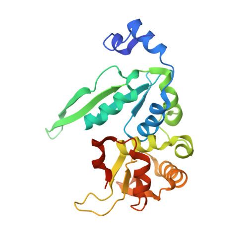
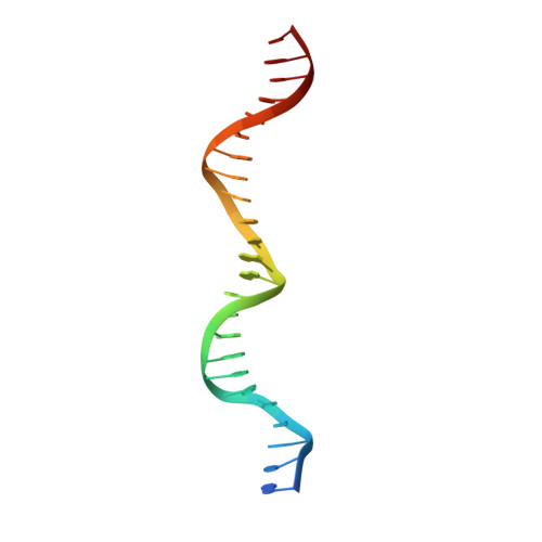
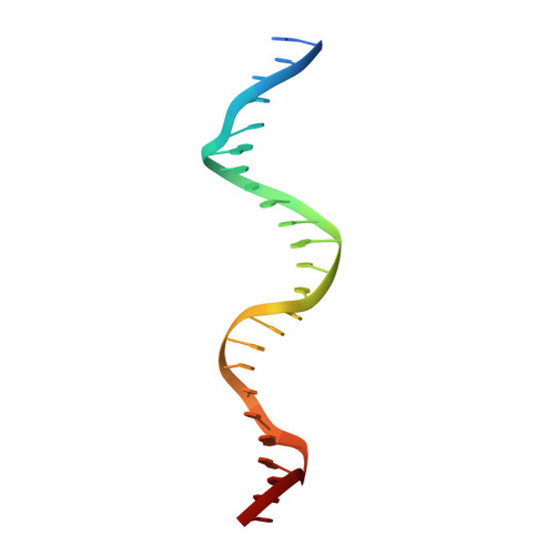
Evolution of I-SceI Homing Endonucleases with Increased DNA Recognition Site Specificity.

Joshi, R.Ho, K.K.Tenney, K.Chen, J.H.Golden, B.L.Gimble, F.S.

(2011) J Mol Biol 405: 185-200

  • DOI: https://doi.org/10.1016/j.jmb.2010.10.029
  • Primary Citation of Related Structures:  
    3OOL, 3OOR

  • PubMed Abstract: 

    Elucidating how homing endonucleases undergo changes in recognition site specificity will facilitate efforts to engineer proteins for gene therapy applications. I-SceI is a monomeric homing endonuclease that recognizes and cleaves within an 18-bp target. It tolerates limited degeneracy in its target sequence, including substitution of a C:G(+4) base pair for the wild-type A:T(+4) base pair. Libraries encoding randomized amino acids at I-SceI residue positions that contact or are proximal to A:T(+4) were used in conjunction with a bacterial one-hybrid system to select I-SceI derivatives that bind to recognition sites containing either the A:T(+4) or the C:G(+4) base pairs. As expected, isolates encoding wild-type residues at the randomized positions were selected using either target sequence. All I-SceI proteins isolated using the C:G(+4) recognition site included small side-chain substitutions at G100 and either contained (K86R/G100T, K86R/G100S and K86R/G100C) or lacked (G100A, G100T) a K86R substitution. Interestingly, the binding affinities of the selected variants for the wild-type A:T(+4) target are 4- to 11-fold lower than that of wild-type I-SceI, whereas those for the C:G(+4) target are similar. The increased specificity of the mutant proteins is also evident in binding experiments in vivo. These differences in binding affinities account for the observed ∼36-fold difference in target preference between the K86R/G100T and wild-type proteins in DNA cleavage assays. An X-ray crystal structure of the K86R/G100T mutant protein bound to a DNA duplex containing the C:G(+4) substitution suggests how sequence specificity of a homing enzyme can increase. This biochemical and structural analysis defines one pathway by which site specificity is augmented for a homing endonuclease.


  • Organizational Affiliation

    Department of Biochemistry, Purdue University, West Lafayette, IN 47907, USA.


Macromolecules

Find similar proteins by:  (by identity cutoff)  |  3D Structure
Entity ID: 1
MoleculeChains Sequence LengthOrganismDetailsImage
Intron encoded endonuclease I-SceI237Saccharomyces cerevisiaeMutation(s): 0 
Gene Names: SceIomega
EC: 3.1
UniProt
Find proteins for P03882 (Saccharomyces cerevisiae (strain ATCC 204508 / S288c))
Explore P03882 
Go to UniProtKB:  P03882
Entity Groups  
Sequence Clusters30% Identity50% Identity70% Identity90% Identity95% Identity100% Identity
UniProt GroupP03882
Sequence Annotations
Expand
  • Reference Sequence

Find similar nucleic acids by:  Sequence   |   3D Structure  

Entity ID: 2
MoleculeChains LengthOrganismImage
5'-D(*CP*AP*CP*GP*CP*TP*AP*GP*GP*GP*AP*TP*AP*AP*CP*CP*GP*GP*GP*TP*AP*AP*TP*AP*C)-3'B [auth C]25N/A
Sequence Annotations
Expand
  • Reference Sequence

Find similar nucleic acids by:  Sequence   |   3D Structure  

Entity ID: 3
MoleculeChains LengthOrganismImage
5'-D(*GP*GP*TP*AP*TP*TP*AP*CP*CP*CP*GP*GP*TP*TP*AP*TP*CP*CP*CP*TP*AP*GP*CP*GP*T)-3'C [auth D]25N/A
Sequence Annotations
Expand
  • Reference Sequence
Experimental Data & Validation

Experimental Data

  • Method: X-RAY DIFFRACTION
  • Resolution: 2.30 Å
  • R-Value Free: 0.294 
  • R-Value Work: 0.246 
  • R-Value Observed: 0.250 
  • Space Group: C 1 2 1
Unit Cell:
Length ( Å )Angle ( ˚ )
a = 129.184α = 90
b = 49.938β = 107.54
c = 63.36γ = 90
Software Package:
Software NamePurpose
DENZOdata reduction
SCALEPACKdata scaling
REFMACrefinement
PDB_EXTRACTdata extraction
HKL-2000data collection

Structure Validation

View Full Validation Report


Entry History 

Deposition Data

Revision History  (Full details and data files)

  • Version 1.0: 2010-11-17
    Type: Initial release
  • Version 1.1: 2011-07-13
    Changes: Version format compliance
  • Version 1.2: 2017-11-08
    Changes: Refinement description
  • Version 1.3: 2018-01-24
    Changes: Structure summary
  • Version 1.4: 2023-09-06
    Changes: Data collection, Database references, Derived calculations, Refinement description