Crystal and solution structures of an odorant-binding protein from the southern house mosquito complexed with an oviposition pheromone.
Mao, Y., Xu, X., Xu, W., Ishida, Y., Leal, W.S., Ames, J.B., Clardy, J.(2010) Proc Natl Acad Sci U S A 107: 19102-19107
- PubMed: 20956299
- DOI: https://doi.org/10.1073/pnas.1012274107
- Primary Citation of Related Structures:
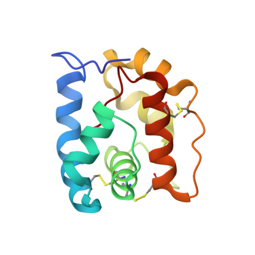
3OGN - PubMed Abstract:
Culex mosquitoes introduce the pathogens responsible for filariasis, West Nile virus, St. Louis encephalitis, and other diseases into humans. Currently, traps baited with oviposition semiochemicals play an important role in detection efforts and could provide an environmentally friendly approach to controlling their populations. The odorant binding proteins (OBPs) in the female's antenna play a crucial, if yet imperfectly understood, role in sensing oviposition cues. Here, we report the X-ray crystallography and NMR 3D structures of OBP1 for Culex quinquefasciatus (CquiOBP1) bound to an oviposition pheromone (5R,6S)-6-acetoxy-5-hexadecanolide (MOP). In both studies, CquiOBP1 had the same overall six-helix structure seen in other insect OBPs, but a detailed analysis revealed an important previously undescribed feature. There are two models for OBP-mediated signal transduction: (i) direct release of the pheromone from an internal binding pocket in a pH-dependent fashion and (ii) detection of a pheromone-induced conformational change in the OBP·pheromone complex. Although CquiOBP1 binds MOP in a pH-dependent fashion, it lacks the C terminus required for the pH-dependent release model. This study shows that CquiOBP binds MOP in an unprecedented fashion using both a small central cavity for the lactone head group and a long hydrophobic channel for its tail.
Organizational Affiliation:
Department of Biological Chemistry and Molecular Pharmacology, Harvard Medical School, Boston, MA 02115, USA.