Crystal Structures of NodS N-Methyltransferase from Bradyrhizobium japonicum in Ligand-Free Form and as SAH Complex.
Cakici, O., Sikorski, M., Stepkowski, T., Bujacz, G., Jaskolski, M.(2010) J Mol Biol 404: 874-889
- PubMed: 20970431
- DOI: https://doi.org/10.1016/j.jmb.2010.10.016
- Primary Citation of Related Structures:
3OFJ, 3OFK - PubMed Abstract:
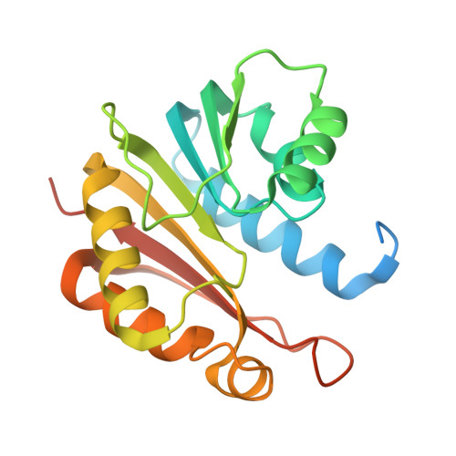
NodS is an S-adenosyl-L-methionine (SAM)-dependent N-methyltransferase that is involved in the biosynthesis of Nod factor (NF) in rhizobia, which are bacterial symbionts of legume plants. NF is a modified chitooligosaccharide (COS) signal molecule that is recognized by the legume host, where it initiates symbiotic processes leading to atmospheric nitrogen fixation. We report the crystal structure of recombinant NodS protein from Bradyrhizobium japonicum, which infects lupine and serradella legumes. Two crystal forms--ligand-free NodS and NodS in complex with S-adenosyl-L-homocysteine, which is a by-product of the methylation reaction--were obtained, and their structures were refined to resolutions of 2.43 Å and 1.85 Å, respectively. Although the overall fold (consisting of a seven-stranded β-sheet flanked by layers of helices) is similar to those of other SAM-dependent methyltransferases, NodS has specific features reflecting the unique character of its oligosaccharide substrate. In particular, the N-terminal helix and its connecting loop get ordered upon SAM binding, thereby closing the methyl donor cavity and shaping a long surface canyon that is clearly the binding site for the acceptor molecule. Comparison of the two structural forms of NodS suggests that there are also other conformational changes taking place upon the binding of the donor substrate. As an enzyme that methylates a COS substrate, NodS is the first example among all SAM-dependent methyltransferases to have its three-dimensional structure elucidated. Gaining insight about how NodS binds its donor and acceptor substrates helps to better understand the mechanism of NodS activity and the basis of its functional difference in various rhizobia.
Organizational Affiliation:
Institute of Bioorganic Chemistry, Polish Academy of Sciences, Poznan, Poland.