3OED

The structure of the complex between complement receptor CR2 and its ligand complement fragment C3d


Experimental Data Snapshot

  • Method: X-RAY DIFFRACTION
  • Resolution: 3.16 Å
  • R-Value Free: 0.259 
  • R-Value Work: 0.189 

wwPDB Validation   3D Report Full Report


This is version 1.3 of the entry. See complete history


Literature

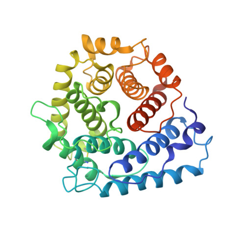
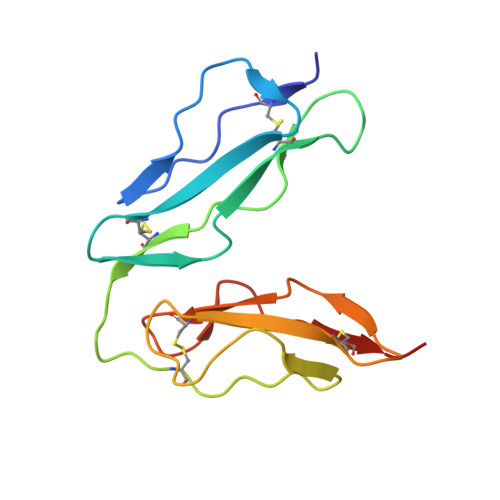
A crystal structure of the complex between human complement receptor 2 and its ligand C3d.

van den Elsen, J.M.Isenman, D.E.

(2011) Science 332: 608-611

  • DOI: https://doi.org/10.1126/science.1201954
  • Primary Citation of Related Structures:  
    3OED

  • PubMed Abstract: 

    The interaction of complement receptor 2 (CR2)--which is present on B cells and follicular dendritic cells--with its antigen-bound ligand C3d results in an enhanced antibody response, thus providing an important link between the innate and adaptive immune systems. Although a cocrystal structure of a complex between C3d and the ligand-binding domains of CR2 has been published, several aspects of this structure, including the position in C3d of the binding interface, remained controversial because of disagreement with biochemical data. We now report a cocrystal structure of a CR2(SCR1-2):C3d complex at 3.2 angstrom resolution in which the interaction interfaces differ markedly from the previously published structure and are consistent with the biochemical data. It is likely that, in the previous structure, the interaction was influenced by the presence of zinc acetate additive in the crystallization buffer, leading to a nonphysiological complex. Detailed knowledge of the binding interface now at hand gives the potential to exploit the interaction in vaccine design or in therapeutics directed against autoreactive B cells.


  • Organizational Affiliation

    Department of Biology and Biochemistry, University of Bath, Bath BA2 7AY, UK. bssjmhve@bath.ac.uk


Macromolecules
Find similar proteins by:  (by identity cutoff)  |  3D Structure
Entity ID: 1
MoleculeChains Sequence LengthOrganismDetailsImage
Complement C3A,
C [auth B]
310Homo sapiensMutation(s): 1 
Gene Names: C3CPAMD1
UniProt & NIH Common Fund Data Resources
Find proteins for P01024 (Homo sapiens)
Explore P01024 
Go to UniProtKB:  P01024
PHAROS:  P01024
GTEx:  ENSG00000125730 
Entity Groups  
Sequence Clusters30% Identity50% Identity70% Identity90% Identity95% Identity100% Identity
UniProt GroupP01024
Sequence Annotations
Expand
  • Reference Sequence
Find similar proteins by:  (by identity cutoff)  |  3D Structure
Entity ID: 2
MoleculeChains Sequence LengthOrganismDetailsImage
Complement receptor type 2B [auth C],
D
135Homo sapiensMutation(s): 0 
Gene Names: C3DRCR2
UniProt & NIH Common Fund Data Resources
Find proteins for P20023 (Homo sapiens)
Explore P20023 
Go to UniProtKB:  P20023
PHAROS:  P20023
GTEx:  ENSG00000117322 
Entity Groups  
Sequence Clusters30% Identity50% Identity70% Identity90% Identity95% Identity100% Identity
UniProt GroupP20023
Sequence Annotations
Expand
  • Reference Sequence
Experimental Data & Validation

Experimental Data

  • Method: X-RAY DIFFRACTION
  • Resolution: 3.16 Å
  • R-Value Free: 0.259 
  • R-Value Work: 0.189 
  • Space Group: H 3 2
Unit Cell:
Length ( Å )Angle ( ˚ )
a = 146.12α = 90
b = 146.12β = 90
c = 253.06γ = 120
Software Package:
Software NamePurpose
ADSCdata collection
BALBESphasing
REFMACrefinement
HKL-2000data reduction
HKL-2000data scaling

Structure Validation

View Full Validation Report


Entry History 

Deposition Data

Revision History  (Full details and data files)

  • Version 1.0: 2011-05-11
    Type: Initial release
  • Version 1.1: 2011-07-13
    Changes: Version format compliance
  • Version 1.2: 2012-03-28
    Changes: Database references
  • Version 1.3: 2023-09-06
    Changes: Data collection, Database references, Refinement description