3ODH

Structure of OkrAI/DNA complex


Experimental Data Snapshot

  • Method: X-RAY DIFFRACTION
  • Resolution: 2.30 Å
  • R-Value Free: 0.234 
  • R-Value Work: 0.155 
  • R-Value Observed: 0.159 

wwPDB Validation   3D Report Full Report


This is version 1.3 of the entry. See complete history


Literature

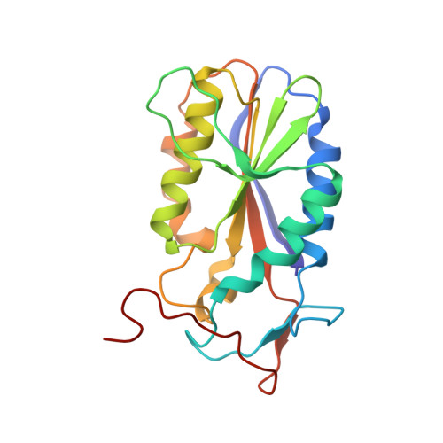
Asymmetric DNA recognition by the OkrAI endonuclease, an isoschizomer of BamHI.

Vanamee, E.S.Viadiu, H.Chan, S.H.Ummat, A.Hartline, A.M.Xu, S.Y.Aggarwal, A.K.

(2011) Nucleic Acids Res 39: 712-719

  • DOI: https://doi.org/10.1093/nar/gkq779
  • Primary Citation of Related Structures:  
    3ODH

  • PubMed Abstract: 

    Restriction enzymes share little or no sequence homology with the exception of isoschizomers, or enzymes that recognize and cleave the same DNA sequence. We present here the structure of a BamHI isoschizomer, OkrAI, bound to the same DNA sequence (TATGGATCCATA) as that cocrystallized with BamHI. We show that OkrAI is a more minimal version of BamHI, lacking not only the N- and C-terminal helices but also an internal 3(10) helix and containing β-strands that are shorter than those in BamHI. Despite these structural differences, OkrAI recognizes the DNA in a remarkably similar manner to BamHI, including asymmetric contacts via C-terminal 'arms' that appear to 'compete' for the minor groove. However, the arms are shorter than in BamHI. We observe similar DNA-binding affinities between OkrAI and BamHI but OkrAI has higher star activity (at 37°C) compared to BamHI. Together, the OkrAI and BamHI structures offer a rare opportunity to compare two restriction enzymes that work on exactly the same DNA substrate.


  • Organizational Affiliation

    Department of Structural and Chemical Biology, Mount Sinai School of Medicine, New York, NY 10029, USA.


Macromolecules

Find similar proteins by:  (by identity cutoff)  |  3D Structure
Entity ID: 1
MoleculeChains Sequence LengthOrganismDetailsImage
OkrAI endonucleaseA,
B,
C [auth E],
D [auth F]
194Oceanobacter kriegiiMutation(s): 0 
Entity Groups  
Sequence Clusters30% Identity50% Identity70% Identity90% Identity95% Identity100% Identity
Sequence Annotations
Expand
  • Reference Sequence

Find similar nucleic acids by:  Sequence   |   3D Structure  

Entity ID: 2
MoleculeChains LengthOrganismImage
DNA (5'-D(*TP*AP*TP*GP*GP*AP*TP*CP*CP*AP*TP*A)-3')E [auth C],
F [auth D],
G,
H
12N/A
Sequence Annotations
Expand
  • Reference Sequence
Experimental Data & Validation

Experimental Data

  • Method: X-RAY DIFFRACTION
  • Resolution: 2.30 Å
  • R-Value Free: 0.234 
  • R-Value Work: 0.155 
  • R-Value Observed: 0.159 
  • Space Group: P 21 21 21
Unit Cell:
Length ( Å )Angle ( ˚ )
a = 80.982α = 90
b = 83.075β = 90
c = 119.67γ = 90
Software Package:
Software NamePurpose
REFMACrefinement
CNSrefinement
DENZOdata reduction
HKL-2000data scaling
CNSphasing

Structure Validation

View Full Validation Report


Entry History 

Deposition Data

Revision History  (Full details and data files)

  • Version 1.0: 2010-10-06
    Type: Initial release
  • Version 1.1: 2011-07-13
    Changes: Version format compliance
  • Version 1.2: 2019-07-17
    Changes: Data collection, Refinement description
  • Version 1.3: 2024-02-21
    Changes: Data collection, Database references, Derived calculations