3O3U

Crystal Structure of Human Receptor for Advanced Glycation Endproducts (RAGE)


Experimental Data Snapshot

  • Method: X-RAY DIFFRACTION
  • Resolution: 1.50 Å
  • R-Value Free: 0.184 
  • R-Value Work: 0.168 
  • R-Value Observed: 0.169 

wwPDB Validation   3D Report Full Report


This is version 2.0 of the entry. See complete history


Literature

The 1.5 A crystal structure of human receptor for advanced glycation endproducts (RAGE) ectodomains reveals unique features determining ligand binding.

Park, H.Boyington, J.C.

(2010) J Biol Chem 285: 40762-40770

  • DOI: https://doi.org/10.1074/jbc.M110.169276
  • Primary Citation of Related Structures:  
    3O3U

  • PubMed Abstract: 

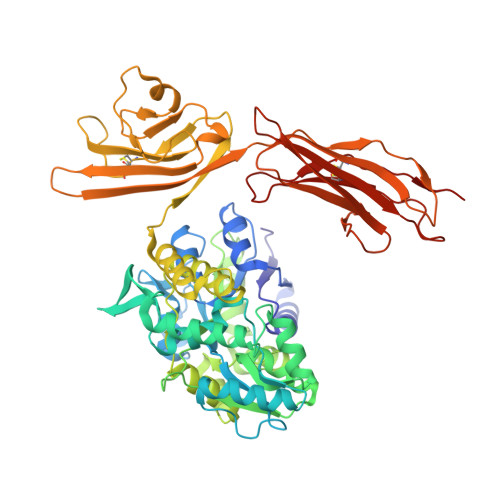
    Interaction of the pattern recognition receptor, RAGE with key ligands such as advanced glycation end products (AGE), S100 proteins, amyloid β, and HMGB1 has been linked to diabetic complications, inflammatory and neurodegenerative disorders, and cancer. To help answer the question of how a single receptor can recognize and respond to a diverse set of ligands we have investigated the structure and binding properties of the first two extracellular domains of human RAGE, which are implicated in various ligand binding and subsequent signaling events. The 1.5-Å crystal structure reveals an elongated molecule with a large basic patch and a large hydrophobic patch, both highly conserved. Isothermal titration calorimetry (ITC) and deletion experiments indicate S100B recognition by RAGE is an entropically driven process involving hydrophobic interaction that is dependent on Ca(2+) and on residues in the C'D loop (residues 54-67) of domain 1. In contrast, competition experiments using gel shift assays suggest that RAGE interaction with AGE is driven by the recognition of negative charges on AGE-proteins. We also demonstrate that RAGE can bind to dsDNA and dsRNA. These findings reveal versatile structural features of RAGE that help explain its ability to recognize of multiple ligands.


  • Organizational Affiliation

    Laboratory of Structural Biology, NIEHS, National Institutes of Health, Research Triangle Park, North Carolina 27709, USA. hajpark@scripps.edu


Macromolecules
Find similar proteins by:  (by identity cutoff)  |  3D Structure
Entity ID: 1
MoleculeChains Sequence LengthOrganismDetailsImage
Maltose-binding periplasmic protein, Advanced glycosylation end product-specific receptorA [auth N]581Escherichia coliHomo sapiens
This entity is chimeric
Mutation(s): 0 
Gene Names: b4034JW3994malErageAGER
UniProt & NIH Common Fund Data Resources
Find proteins for P0AEX9 (Escherichia coli (strain K12))
Explore P0AEX9 
Go to UniProtKB:  P0AEX9
Find proteins for Q15109 (Homo sapiens)
Explore Q15109 
Go to UniProtKB:  Q15109
PHAROS:  Q15109
GTEx:  ENSG00000204305 
Entity Groups  
Sequence Clusters30% Identity50% Identity70% Identity90% Identity95% Identity100% Identity
UniProt GroupsQ15109P0AEX9
Sequence Annotations
Expand
  • Reference Sequence
Oligosaccharides

Help

Entity ID: 2
MoleculeChains Length2D Diagram Glycosylation3D Interactions
alpha-D-glucopyranose-(1-4)-alpha-D-glucopyranose-(1-4)-alpha-D-glucopyranoseB [auth A]3N/A
Glycosylation Resources
GlyTouCan:  G96370VA
GlyCosmos:  G96370VA
GlyGen:  G96370VA
Small Molecules
Ligands 1 Unique
IDChains Name / Formula / InChI Key2D Diagram3D Interactions
SO4
Query on SO4

Download Ideal Coordinates CCD File 
C [auth N]SULFATE ION
O4 S
QAOWNCQODCNURD-UHFFFAOYSA-L
Biologically Interesting Molecules (External Reference) 1 Unique
Experimental Data & Validation

Experimental Data

  • Method: X-RAY DIFFRACTION
  • Resolution: 1.50 Å
  • R-Value Free: 0.184 
  • R-Value Work: 0.168 
  • R-Value Observed: 0.169 
  • Space Group: P 21 21 2
Unit Cell:
Length ( Å )Angle ( ˚ )
a = 81.739α = 90
b = 89.308β = 90
c = 97.978γ = 90
Software Package:
Software NamePurpose
SERGUIdata collection
MOLREPphasing
PHENIXrefinement
HKL-2000data reduction
HKL-2000data scaling

Structure Validation

View Full Validation Report


Entry History 

Deposition Data

Revision History  (Full details and data files)

  • Version 1.0: 2010-10-13
    Type: Initial release
  • Version 1.1: 2011-07-13
    Changes: Version format compliance
  • Version 1.2: 2017-08-02
    Changes: Advisory, Refinement description, Source and taxonomy
  • Version 2.0: 2020-07-29
    Type: Remediation
    Reason: Carbohydrate remediation
    Changes: Atomic model, Data collection, Database references, Derived calculations, Non-polymer description, Structure summary