3NUB

WbpE, an Aminotransferase from Pseudomonas aeruginosa Involved in O-antigen Assembly in Complex with Product as the External Aldimine


Experimental Data Snapshot

  • Method: X-RAY DIFFRACTION
  • Resolution: 1.90 Å
  • R-Value Free: 0.243 
  • R-Value Work: 0.199 
  • R-Value Observed: 0.202 

wwPDB Validation   3D Report Full Report


Ligand Structure Quality Assessment 


This is version 1.2 of the entry. See complete history


Literature

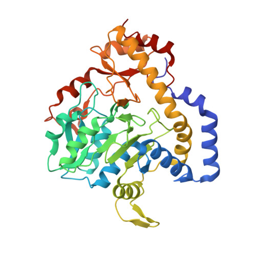
Structural Analysis of WbpE from Pseudomonas aeruginosa PAO1: A Nucleotide Sugar Aminotransferase Involved in O-Antigen Assembly

Larkin, A.Olivier, N.B.Imperiali, B.

(2010) Biochemistry 49: 7227-7237

  • DOI: https://doi.org/10.1021/bi100805b
  • Primary Citation of Related Structures:  
    3NU7, 3NU8, 3NUB

  • PubMed Abstract: 

    In recent years, the opportunistic pathogen Pseudomonas aeruginosa has emerged as a major source of hospital-acquired infections. Effective treatment has proven increasingly difficult due to the spread of multidrug resistant strains and thus requires a deeper understanding of the biochemical mechanisms of pathogenicity. The central carbohydrate of the P. aeruginosa PAO1 (O5) B-band O-antigen, ManNAc(3NAc)A, has been shown to be critical for virulence and is produced in a stepwise manner by five enzymes in the Wbp pathway (WbpA, WbpB, WbpE, WbpD, and WbpI). Herein, we present the crystal structure of the aminotransferase WbpE from P. aeruginosa PAO1 in complex with the cofactor pyridoxal 5'-phosphate (PLP) and product UDP-GlcNAc(3NH(2))A as the external aldimine at 1.9 A resolution. We also report the structures of WbpE in complex with PMP alone as well as the PLP internal aldimine and show that the dimeric structure of WbpE observed in the crystal structure is confirmed by analytical ultracentrifugation. Analysis of these structures reveals that the active site of the enzyme is composed of residues from both subunits. In particular, we show that a key residue (Arg229), which has previously been implicated in direct interactions with the alpha-carboxylate moiety of alpha-ketoglutarate, is also uniquely positioned to bestow specificity for the 6''-carboxyl group of GlcNAc(3NH(2))A through a salt bridge. This finding is intriguing because while an analogous basic residue is present in WbpE homologues that do not process 6''-carboxyl-modified saccharides, recent structural studies reveal that this side chain is retracted to accommodate a neutral C6'' atom. This work represents the first structural analysis of a nucleotide sugar aminotransferase with a bound product modified at the C2'', C3'', and C6'' positions and provides insight into a novel target for treatment of P. aeruginosa infection.


  • Organizational Affiliation

    Department of Chemistry, Massachusetts Institute of Technology, 77 Massachusetts Avenue, Cambridge, Massachusetts 02139, USA.


Macromolecules
Find similar proteins by:  (by identity cutoff)  |  3D Structure
Entity ID: 1
MoleculeChains Sequence LengthOrganismDetailsImage
Aminotransferase WbpE
A, B
359Pseudomonas aeruginosaMutation(s): 0 
Gene Names: PA3155wbpE
UniProt
Find proteins for Q9HZ76 (Pseudomonas aeruginosa (strain ATCC 15692 / DSM 22644 / CIP 104116 / JCM 14847 / LMG 12228 / 1C / PRS 101 / PAO1))
Explore Q9HZ76 
Go to UniProtKB:  Q9HZ76
Entity Groups  
Sequence Clusters30% Identity50% Identity70% Identity90% Identity95% Identity100% Identity
UniProt GroupQ9HZ76
Sequence Annotations
Expand
  • Reference Sequence
Small Molecules
Ligands 3 Unique
IDChains Name / Formula / InChI Key2D Diagram3D Interactions
UD0
Query on UD0

Download Ideal Coordinates CCD File 
C [auth A],
F [auth B]
(2S,3S,4R,5R,6R)-5-(acetylamino)-6-{[(S)-{[(S)-{[(2S,3R,4S,5R)-5-(2,4-dioxo-3,4-dihydropyrimidin-1(2H)-yl)-3,4-dihydroxytetrahydrofuran-2-yl]methoxy}(hydroxy)phosphoryl]oxy}(hydroxy)phosphoryl]oxy}-3-hydroxy-4-{[(1E)-{3-hydroxy-2-methyl-5-[(phosphonooxy)methyl]pyridin-4-yl}methylidene]amino}tetrahydro-2H-pyran-2-carboxylic acid
C25 H34 N5 O22 P3
IGKBPIFLZAFLLV-VUZHIUKYSA-N
SO4
Query on SO4

Download Ideal Coordinates CCD File 
E [auth A]SULFATE ION
O4 S
QAOWNCQODCNURD-UHFFFAOYSA-L
GOL
Query on GOL

Download Ideal Coordinates CCD File 
D [auth A]GLYCEROL
C3 H8 O3
PEDCQBHIVMGVHV-UHFFFAOYSA-N
Experimental Data & Validation

Experimental Data

  • Method: X-RAY DIFFRACTION
  • Resolution: 1.90 Å
  • R-Value Free: 0.243 
  • R-Value Work: 0.199 
  • R-Value Observed: 0.202 
  • Space Group: P 21 21 2
Unit Cell:
Length ( Å )Angle ( ˚ )
a = 77.924α = 90
b = 148.679β = 90
c = 53.545γ = 90
Software Package:
Software NamePurpose
HKL-2000data collection
PHASERphasing
REFMACrefinement
HKL-2000data reduction
HKL-2000data scaling

Structure Validation

View Full Validation Report


Ligand Structure Quality Assessment 


Entry History 

Deposition Data

Revision History  (Full details and data files)

  • Version 1.0: 2010-07-28
    Type: Initial release
  • Version 1.1: 2011-07-13
    Changes: Version format compliance
  • Version 1.2: 2023-09-06
    Changes: Data collection, Database references, Derived calculations, Refinement description