3NR2

Crystal structure of Caspase-6 zymogen


Experimental Data Snapshot

  • Method: X-RAY DIFFRACTION
  • Resolution: 2.90 Å
  • R-Value Free: 0.237 
  • R-Value Work: 0.179 
  • R-Value Observed: 0.185 

wwPDB Validation   3D Report Full Report


This is version 1.2 of the entry. See complete history


Literature

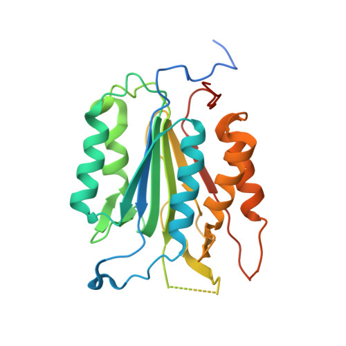
Crystal structures of human caspase 6 reveal a new mechanism for intramolecular cleavage self-activation

Wang, X.-J.Cao, Q.Liu, X.Wang, K.-T.Mi, W.Zhang, Y.Li, L.-F.Leblanc, A.C.Su, X.-D.

(2010) EMBO Rep 11: 841-847

  • DOI: https://doi.org/10.1038/embor.2010.141
  • Primary Citation of Related Structures:  
    3NR2, 3OD5

  • PubMed Abstract: 

    Dimeric effectors caspase 3 and caspase 7 are activated by initiator caspase processing. In this study, we report the crystal structures of effector caspase 6 (CASP6) zymogen and N-Acetyl-Val-Glu-Ile-Asp-al-inhibited CASP6. Both of these forms of CASP6 have a dimeric structure, and in CASP6 zymogen the intersubunit cleavage site (190)TEVD(193) is well structured and inserts into the active site. This positions residue Asp 193 to be easily attacked by the catalytic residue Cys 163. We demonstrate biochemically that intramolecular cleavage at Asp 193 is a prerequisite for CASP6 self-activation and that this activation mechanism is dependent on the length of the L2 loop. Our results indicate that CASP6 can be activated and regulated through intramolecular self-cleavage.


  • Organizational Affiliation

    National Laboratory of Protein Engineering & Plant Genetic Engineering, Peking University, Haidian District, Beijing, People's Republic of China.


Macromolecules
Find similar proteins by:  (by identity cutoff)  |  3D Structure
Entity ID: 1
MoleculeChains Sequence LengthOrganismDetailsImage
Caspase-6
A, B
294Homo sapiensMutation(s): 2 
EC: 3.4.22.59
UniProt & NIH Common Fund Data Resources
Find proteins for P55212 (Homo sapiens)
Explore P55212 
Go to UniProtKB:  P55212
PHAROS:  P55212
GTEx:  ENSG00000138794 
Entity Groups  
Sequence Clusters30% Identity50% Identity70% Identity90% Identity95% Identity100% Identity
UniProt GroupP55212
Sequence Annotations
Expand
  • Reference Sequence
Experimental Data & Validation

Experimental Data

  • Method: X-RAY DIFFRACTION
  • Resolution: 2.90 Å
  • R-Value Free: 0.237 
  • R-Value Work: 0.179 
  • R-Value Observed: 0.185 
  • Space Group: P 65 2 2
Unit Cell:
Length ( Å )Angle ( ˚ )
a = 127.983α = 90
b = 127.983β = 90
c = 167.912γ = 120
Software Package:
Software NamePurpose
PROTEUM PLUSdata collection
MOLREPphasing
PHENIXrefinement
PROTEUM PLUSdata reduction
PROTEUM PLUSdata scaling

Structure Validation

View Full Validation Report


Entry History 

Deposition Data

Revision History  (Full details and data files)

  • Version 1.0: 2010-10-27
    Type: Initial release
  • Version 1.1: 2011-07-13
    Changes: Version format compliance
  • Version 1.2: 2023-11-01
    Changes: Data collection, Database references, Refinement description