3NKU

Crystal structure of the N-terminal domain of DrrA/SidM from Legionella pneumophila


Experimental Data Snapshot

  • Method: X-RAY DIFFRACTION
  • Resolution: 2.10 Å
  • R-Value Free: 0.228 
  • R-Value Work: 0.180 
  • R-Value Observed: 0.182 

wwPDB Validation   3D Report Full Report


This is version 1.1 of the entry. See complete history


Literature

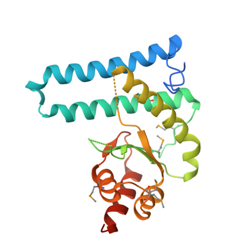
The Legionella effector protein DrrA AMPylates the membrane traffic regulator Rab1b.

Muller, M.P.Peters, H.Blumer, J.Blankenfeldt, W.Goody, R.S.Itzen, A.

(2010) Science 329: 946-949

  • DOI: https://doi.org/10.1126/science.1192276
  • Primary Citation of Related Structures:  
    3NKU, 3NKV

  • PubMed Abstract: 

    In the course of Legionnaires' disease, the bacterium Legionella pneumophila affects the intracellular vesicular trafficking of infected eukaryotic cells by recruiting the small guanosine triphosphatase (GTPase) Rab1 to the cytosolic face of the Legionella-containing vacuole. In order to accomplish this, the Legionella protein DrrA contains a specific guanine nucleotide exchange activity for Rab1 activation that exchanges guanosine triphosphate (GTP) for guanosine diphosphate on Rab1. We found that the amino-terminal domain of DrrA possesses adenosine monophosphorylation (AMPylation) activity toward the switch II region of Rab1b, leading to posttranslational covalent modification of tyrosine 77. AMPylation of switch II by DrrA restricts the access of GTPase activating proteins, thereby rendering Rab1b constitutively active.


  • Organizational Affiliation

    Department of Physical Biochemistry, Max Planck Institute of Molecular Physiology, Dortmund, NRW, 44227, Germany.


Macromolecules
Find similar proteins by:  (by identity cutoff)  |  3D Structure
Entity ID: 1
MoleculeChains Sequence LengthOrganismDetailsImage
DrrA
A, B
213Legionella pneumophila subsp. pneumophila str. Philadelphia 1Mutation(s): 0 
Gene Names: DrrA
UniProt
Find proteins for Q29ST3 (Legionella pneumophila)
Explore Q29ST3 
Go to UniProtKB:  Q29ST3
Entity Groups  
Sequence Clusters30% Identity50% Identity70% Identity90% Identity95% Identity100% Identity
UniProt GroupQ29ST3
Sequence Annotations
Expand
  • Reference Sequence
Small Molecules
Ligands 2 Unique
IDChains Name / Formula / InChI Key2D Diagram3D Interactions
PGE
Query on PGE

Download Ideal Coordinates CCD File 
D [auth A]TRIETHYLENE GLYCOL
C6 H14 O4
ZIBGPFATKBEMQZ-UHFFFAOYSA-N
PEG
Query on PEG

Download Ideal Coordinates CCD File 
C [auth A]DI(HYDROXYETHYL)ETHER
C4 H10 O3
MTHSVFCYNBDYFN-UHFFFAOYSA-N
Modified Residues  1 Unique
IDChains TypeFormula2D DiagramParent
MSE
Query on MSE
A, B
L-PEPTIDE LINKINGC5 H11 N O2 SeMET
Experimental Data & Validation

Experimental Data

  • Method: X-RAY DIFFRACTION
  • Resolution: 2.10 Å
  • R-Value Free: 0.228 
  • R-Value Work: 0.180 
  • R-Value Observed: 0.182 
  • Space Group: P 21 21 21
Unit Cell:
Length ( Å )Angle ( ˚ )
a = 47.609α = 90
b = 49.955β = 90
c = 159.775γ = 90
Software Package:
Software NamePurpose
XSCALEdata scaling
REFMACrefinement
PDB_EXTRACTdata extraction
XDSdata reduction

Structure Validation

View Full Validation Report


Entry History 

Deposition Data

Revision History  (Full details and data files)

  • Version 1.0: 2010-08-04
    Type: Initial release
  • Version 1.1: 2011-07-13
    Changes: Version format compliance