


Crystal structure of AP endonuclease, family 2 from Brucella melitensis
Staker, B., Edwards, T., Bullen, J., Dieterich, M., Seattle Structural Genomics Center for Infectious Disease (SSGCID)To be published.
Experimental Data Snapshot
wwPDB Validation 3D Report Full Report

Entity ID: 1 | |||||
|---|---|---|---|---|---|
| Molecule | Chains | Sequence Length | Organism | Details | Image |
| AP endonuclease, family 2 | 269 | Brucella abortus 2308 | Mutation(s): 0 Gene Names: BAB2_0144 EC: 5.3.1.22 | 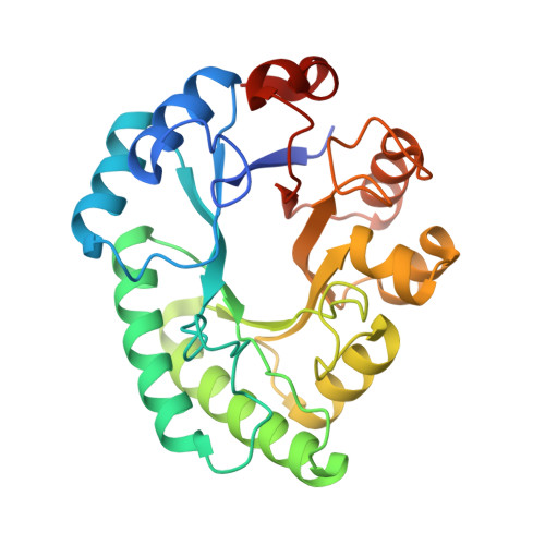 | |
UniProt | |||||
Find proteins for Q2YLA2 (Brucella abortus (strain 2308)) Explore Q2YLA2 Go to UniProtKB: Q2YLA2 | |||||
Entity Groups | |||||
| Sequence Clusters | 30% Identity50% Identity70% Identity90% Identity95% Identity100% Identity | ||||
| UniProt Group | Q2YLA2 | ||||
Sequence AnnotationsExpand | |||||
| |||||
| Ligands 2 Unique | |||||
|---|---|---|---|---|---|
| ID | Chains | Name / Formula / InChI Key | 2D Diagram | 3D Interactions | |
| GOL Query on GOL | D [auth A] E [auth A] F [auth A] H [auth B] I [auth B] | GLYCEROL C3 H8 O3 PEDCQBHIVMGVHV-UHFFFAOYSA-N |  | ||
| MN Query on MN | C [auth A], G [auth B] | MANGANESE (II) ION Mn WAEMQWOKJMHJLA-UHFFFAOYSA-N |  | ||
| Length ( Å ) | Angle ( ˚ ) |
|---|---|
| a = 52.98 | α = 90 |
| b = 120.64 | β = 117.14 |
| c = 54.68 | γ = 90 |
| Software Name | Purpose |
|---|---|
| StructureStudio | data collection |
| PHASER | phasing |
| REFMAC | refinement |
| XDS | data reduction |
| XSCALE | data scaling |
RCSB PDB (citation) is hosted by
RCSB PDB is a member of the

