3NB8

Crystal structure of oxidized H88Q Synechocystis sp. PCYA


Experimental Data Snapshot

  • Method: X-RAY DIFFRACTION
  • Resolution: 1.30 Å
  • R-Value Free: 0.191 
  • R-Value Work: 0.169 
  • R-Value Observed: 0.170 

wwPDB Validation   3D Report Full Report


Ligand Structure Quality Assessment 


This is version 1.3 of the entry. See complete history


Literature

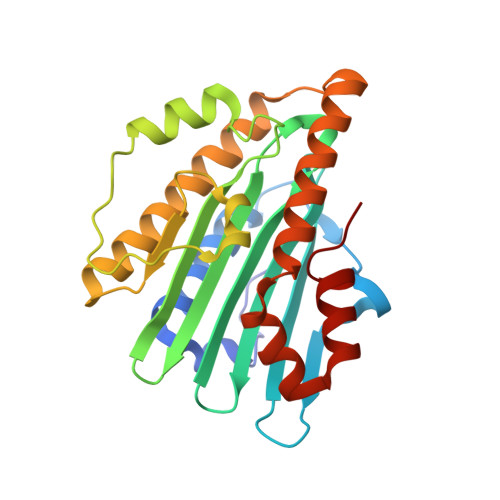
Structural basis for hydration dynamics in radical stabilization of bilin reductase mutants.

Kohler, A.C.Gae, D.D.Richley, M.A.Stoll, S.Gunn, A.Lim, S.Martin, S.S.Doukov, T.I.Britt, R.D.Ames, J.B.Lagarias, J.C.Fisher, A.J.

(2010) Biochemistry 49: 6206-6218

  • DOI: https://doi.org/10.1021/bi100728q
  • Primary Citation of Related Structures:  
    3F0L, 3F0M, 3NB8, 3NB9

  • PubMed Abstract: 

    Heme-derived linear tetrapyrroles (phytobilins) in phycobiliproteins and phytochromes perform critical light-harvesting and light-sensing roles in oxygenic photosynthetic organisms. A key enzyme in their biogenesis, phycocyanobilin:ferredoxin oxidoreductase (PcyA), catalyzes the overall four-electron reduction of biliverdin IXalpha to phycocyanobilin--the common chromophore precursor for both classes of biliproteins. This interconversion occurs via semireduced bilin radical intermediates that are profoundly stabilized by selected mutations of two critical catalytic residues, Asp105 and His88. To understand the structural basis for this stabilization and to gain insight into the overall catalytic mechanism, we report the high-resolution crystal structures of substrate-loaded Asp105Asn and His88Gln mutants of Synechocystis sp. PCC 6803 PcyA in the initial oxidized and one-electron reduced radical states. Unlike wild-type PcyA, both mutants possess a bilin-interacting axial water molecule that is ejected from the active site upon formation of the enzyme-bound neutral radical complex. Structural studies of both mutants also show that the side chain of Glu76 is unfavorably located for D-ring vinyl reduction. On the basis of these structures and companion (15)N-(1)H long-range HMQC NMR analyses to assess the protonation state of histidine residues, we propose a new mechanistic scheme for PcyA-mediated reduction of both vinyl groups of biliverdin wherein an axial water molecule, which prematurely binds and ejects from both mutants upon one electron reduction, is required for catalytic turnover of the semireduced state.


  • Organizational Affiliation

    Department of Chemistry, University of California, One Shields Avenue, Davis, California 95616, USA.


Macromolecules
Find similar proteins by:  (by identity cutoff)  |  3D Structure
Entity ID: 1
MoleculeChains Sequence LengthOrganismDetailsImage
Phycocyanobilin:ferredoxin oxidoreductase248Synechocystis sp. PCC 6803Mutation(s): 1 
Gene Names: pcyAslr0116
EC: 1.3.7.5
UniProt
Find proteins for Q55891 (Synechocystis sp. (strain PCC 6803 / Kazusa))
Explore Q55891 
Go to UniProtKB:  Q55891
Entity Groups  
Sequence Clusters30% Identity50% Identity70% Identity90% Identity95% Identity100% Identity
UniProt GroupQ55891
Sequence Annotations
Expand
  • Reference Sequence
Small Molecules
Ligands 2 Unique
IDChains Name / Formula / InChI Key2D Diagram3D Interactions
BLA
Query on BLA

Download Ideal Coordinates CCD File 
P [auth A]BILIVERDINE IX ALPHA
C33 H34 N4 O6
GWZYPXHJIZCRAJ-SRVCBVSDSA-N
EDO
Query on EDO

Download Ideal Coordinates CCD File 
B [auth A]
C [auth A]
D [auth A]
E [auth A]
F [auth A]
B [auth A],
C [auth A],
D [auth A],
E [auth A],
F [auth A],
G [auth A],
H [auth A],
I [auth A],
J [auth A],
K [auth A],
L [auth A],
M [auth A],
N [auth A],
O [auth A]
1,2-ETHANEDIOL
C2 H6 O2
LYCAIKOWRPUZTN-UHFFFAOYSA-N
Experimental Data & Validation

Experimental Data

  • Method: X-RAY DIFFRACTION
  • Resolution: 1.30 Å
  • R-Value Free: 0.191 
  • R-Value Work: 0.169 
  • R-Value Observed: 0.170 
  • Space Group: P 21 21 2
Unit Cell:
Length ( Å )Angle ( ˚ )
a = 70.785α = 90
b = 95.637β = 90
c = 42.71γ = 90
Software Package:
Software NamePurpose
ADSCdata collection
REFMACrefinement
MOSFLMdata reduction
SCALAdata scaling

Structure Validation

View Full Validation Report


Ligand Structure Quality Assessment 


Entry History 

Deposition Data

  • Released Date: 2010-10-06 
  • Deposition Author(s): Gae, D.D.
  • This entry supersedes: 3F0J

Revision History  (Full details and data files)

  • Version 1.0: 2010-10-06
    Type: Initial release
  • Version 1.1: 2011-07-13
    Changes: Version format compliance
  • Version 1.2: 2015-09-16
    Changes: Database references
  • Version 1.3: 2023-09-06
    Changes: Data collection, Database references, Derived calculations, Refinement description