3N54

Crystal Structure of the GerBC protein


Experimental Data Snapshot

  • Method: X-RAY DIFFRACTION
  • Resolution: 2.30 Å
  • R-Value Free: 0.235 
  • R-Value Work: 0.219 
  • R-Value Observed: 0.219 

wwPDB Validation   3D Report Full Report


This is version 1.2 of the entry. See complete history


Literature

Crystal Structure of the GerBC Component of a Bacillus subtilis Spore Germinant Receptor.

Li, Y.Setlow, B.Setlow, P.Hao, B.

(2010) J Mol Biol 402: 8-16

  • DOI: https://doi.org/10.1016/j.jmb.2010.07.018
  • Primary Citation of Related Structures:  
    3N54

  • PubMed Abstract: 

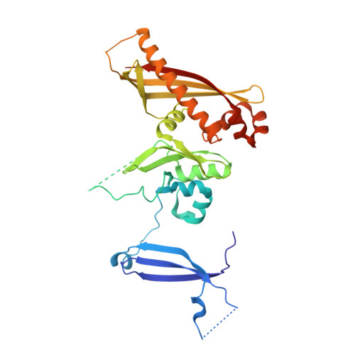
    The nutrient germinant receptors (nGRs) of spores of Bacillus species are clusters of three proteins that play a critical role in triggering the germination of dormant spores in response to specific nutrient molecules. Here, we report the crystal structure of the C protein of the GerB germinant receptor, so-called GerBC, of Bacillus subtilis spores at 2.3 A resolution. The GerBC protein adopts a previously uncharacterized type of protein fold consisting of three distinct domains, each of which is centered by a beta sheet surrounded by multiple alpha helices. Secondary-structure prediction and structure-based sequence alignment suggest that the GerBC structure represents the prototype for C subunits of nGRs from spores of all Bacillales and Clostridiales species and defines two highly conserved structural regions in this family of proteins. GerBC forms an interlocked dimer in the crystalline state but is predominantly monomeric in solution, pointing to the possibility that GerBC oligomerizes as a result of either high local protein concentrations or interaction with other nGR proteins in spores. Our findings provide the first structural view of the nGR subunits and a molecular framework for understanding the architecture, conservation, and function of nGRs.


  • Organizational Affiliation

    Department of Molecular,Microbial and Structural Biology, University of Connecticut Health Center, Farmington, CT 06030-3305, USA.


Macromolecules
Find similar proteins by:  (by identity cutoff)  |  3D Structure
Entity ID: 1
MoleculeChains Sequence LengthOrganismDetailsImage
Spore germination protein B3A [auth B]350Bacillus subtilis subsp. subtilis str. 168Mutation(s): 0 
Gene Names: BSU35820gerBC
UniProt
Find proteins for P39571 (Bacillus subtilis (strain 168))
Explore P39571 
Go to UniProtKB:  P39571
Entity Groups  
Sequence Clusters30% Identity50% Identity70% Identity90% Identity95% Identity100% Identity
UniProt GroupP39571
Sequence Annotations
Expand
  • Reference Sequence
Small Molecules
Ligands 2 Unique
IDChains Name / Formula / InChI Key2D Diagram3D Interactions
SO4
Query on SO4

Download Ideal Coordinates CCD File 
B
C [auth B]
D [auth B]
E [auth B]
F [auth B]
B,
C [auth B],
D [auth B],
E [auth B],
F [auth B],
G [auth B],
H [auth B],
I [auth B],
J [auth B]
SULFATE ION
O4 S
QAOWNCQODCNURD-UHFFFAOYSA-L
CL
Query on CL

Download Ideal Coordinates CCD File 
K [auth B]
L [auth B]
M [auth B]
N [auth B]
O [auth B]
K [auth B],
L [auth B],
M [auth B],
N [auth B],
O [auth B],
P [auth B],
Q [auth B],
R [auth B],
S [auth B]
CHLORIDE ION
Cl
VEXZGXHMUGYJMC-UHFFFAOYSA-M
Experimental Data & Validation

Experimental Data

  • Method: X-RAY DIFFRACTION
  • Resolution: 2.30 Å
  • R-Value Free: 0.235 
  • R-Value Work: 0.219 
  • R-Value Observed: 0.219 
  • Space Group: H 3 2
Unit Cell:
Length ( Å )Angle ( ˚ )
a = 142.785α = 90
b = 142.785β = 90
c = 187.827γ = 120
Software Package:
Software NamePurpose
HKL-2000data collection
PHENIXmodel building
REFMACrefinement
HKL-2000data reduction
HKL-2000data scaling
PHENIXphasing

Structure Validation

View Full Validation Report


Entry History 

Deposition Data

Revision History  (Full details and data files)

  • Version 1.0: 2010-08-04
    Type: Initial release
  • Version 1.1: 2011-07-13
    Changes: Version format compliance
  • Version 1.2: 2024-02-21
    Changes: Data collection, Database references, Derived calculations