3N52

crystal Structure analysis of MIP2


Experimental Data Snapshot

  • Method: X-RAY DIFFRACTION
  • Resolution: 1.90 Å
  • R-Value Free: 0.226 
  • R-Value Work: 0.186 
  • R-Value Observed: 0.188 

wwPDB Validation   3D Report Full Report


This is version 1.4 of the entry. See complete history


Literature

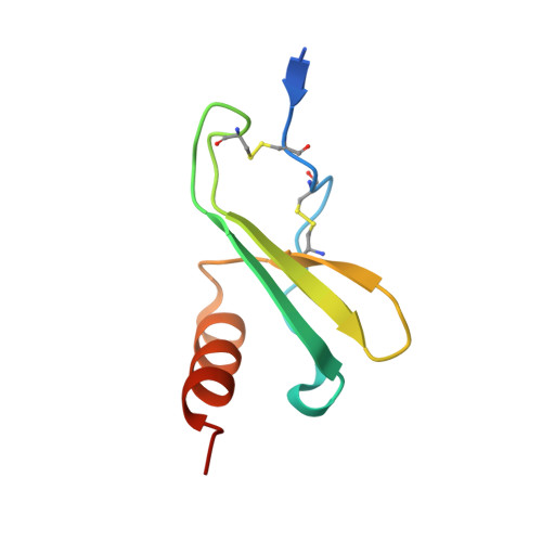
A Model of GAG/MIP-2/CXCR2 Interfaces and Its Functional Effects.

Rajasekaran, D.Keeler, C.Syed, M.A.Jones, M.C.Harrison, J.K.Wu, D.Bhandari, V.Hodsdon, M.E.Lolis, E.J.

(2012) Biochemistry 51: 5642-5654

  • DOI: https://doi.org/10.1021/bi3001566
  • Primary Citation of Related Structures:  
    3N52

  • PubMed Abstract: 

    MIP-2/CXCL2 is a murine chemokine related to human chemokines that possesses the Glu-Leu-Arg (ELR) activation motif and activates CXCR2 for neutrophil chemotaxis. We determined the structure of MIP-2 to 1.9 Å resolution and created a model with its murine receptor CXCR2 based on the coordinates of human CXCR4. Chemokine-induced migration of cells through specific G-protein coupled receptors is regulated by glycosaminoglycans (GAGs) that oligomerize chemokines. MIP-2 GAG-binding residues were identified that interact with heparin disaccharide I-S by NMR spectroscopy. A model GAG/MIP-2/CXCR2 complex that supports a 2:2 complex between chemokine and receptor was created. Mutants of these disaccharide-binding residues were made and tested for heparin binding, in vitro neutrophil chemotaxis, and in vivo neutrophil recruitment to the mouse peritoneum and lung. The mutants have a 10-fold decrease in neutrophil chemotaxis in vitro. There is no difference in neutrophil recruitment between wild-type MIP-2 and mutants in the peritoneum, but all activity of the mutants is lost in the lung, supporting the concept that GAG regulation of chemokines is tissue-dependent.


  • Organizational Affiliation

    Departments of Pharmacology, Yale University School of Medicine, New Haven, CT 06520-8066, USA.


Macromolecules
Find similar proteins by:  (by identity cutoff)  |  3D Structure
Entity ID: 1
MoleculeChains Sequence LengthOrganismDetailsImage
C-X-C motif chemokine 2
A, B, C, D
73Mus musculusMutation(s): 0 
Gene Names: Cxcl2Mip-2Mip2Scyb2
UniProt & NIH Common Fund Data Resources
Find proteins for P10889 (Mus musculus)
Explore P10889 
Go to UniProtKB:  P10889
IMPC:  MGI:1340094
Entity Groups  
Sequence Clusters30% Identity50% Identity70% Identity90% Identity95% Identity100% Identity
UniProt GroupP10889
Sequence Annotations
Expand
  • Reference Sequence
Experimental Data & Validation

Experimental Data

  • Method: X-RAY DIFFRACTION
  • Resolution: 1.90 Å
  • R-Value Free: 0.226 
  • R-Value Work: 0.186 
  • R-Value Observed: 0.188 
  • Space Group: P 21 21 21
Unit Cell:
Length ( Å )Angle ( ˚ )
a = 41.755α = 90
b = 59.446β = 90
c = 99.354γ = 90
Software Package:
Software NamePurpose
DENZOdata reduction
SCALEPACKdata scaling
MOLREPphasing
REFMACrefinement
PDB_EXTRACTdata extraction
CrystalCleardata collection
HKL-2000data reduction

Structure Validation

View Full Validation Report


Entry History 

Deposition Data

Revision History  (Full details and data files)

  • Version 1.0: 2011-06-08
    Type: Initial release
  • Version 1.1: 2011-07-13
    Changes: Version format compliance
  • Version 1.2: 2012-08-01
    Changes: Database references
  • Version 1.3: 2012-10-03
    Changes: Database references
  • Version 1.4: 2017-11-08
    Changes: Refinement description