Crystal structures of bacillus cereus NCTU2 chitinase complexes with chitooligomers reveal novel substrate binding for catalysis: a chitinase without chitin-binding and insertion domains
Hsieh, Y.-C., Wu, Y.-J., Chiang, T.-Y., Kuo, C.-Y., Shrestha, K.L., Chao, C.-F., Huang, Y.-C., Chuankhayan, P., Wu, W.-G., Li, Y.-K., Chen, C.-J.(2010) J Biol Chem 285: 31603-31615
- PubMed: 20685646
- DOI: https://doi.org/10.1074/jbc.M110.149310
- Primary Citation of Related Structures:
3N11, 3N12, 3N13, 3N15, 3N17, 3N18, 3N1A - PubMed Abstract:
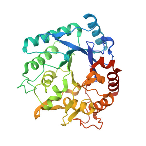
Chitinases hydrolyze chitin, an insoluble linear polymer of N-acetyl-d-glucosamine (NAG)(n), into nutrient sources. Bacillus cereus NCTU2 chitinase (ChiNCTU2) predominantly produces chitobioses and belongs to glycoside hydrolase family 18. The crystal structure of wild-type ChiNCTU2 comprises only a catalytic domain, unlike other chitinases that are equipped with additional chitin binding and insertion domains to bind substrates into the active site. Lacking chitin binding and chitin insertion domains, ChiNCTU2 utilizes two dynamic loops (Gly-67-Thr-69 and Ile-106-Val-112) to interact with (NAG)(n), generating novel substrate binding and distortion for catalysis. Gln-109 is crucial for direct binding with substrates, leading to conformational changes of two loops with a maximum shift of ∼4.6 Å along the binding cleft. The structures of E145Q, E145Q/Y227F, and E145G/Y227F mutants complexed with (NAG)(n) reveal (NAG)(2), (NAG)(2), and (NAG)(4) in the active site, respectively, implying various stages of reaction: before hydrolysis, E145G/Y227F with (NAG)(4); in an intermediate state, E145Q/Y227F with a boat-form NAG at the -1 subsite, -1-(NAG); after hydrolysis, E145Q with a chair form -1-(NAG). Several residues were confirmed to play catalytic roles: Glu-145 in cleavage of the glycosidic bond between -1-(NAG) and +1-(NAG); Tyr-227 in the conformational change of -1-(NAG); Asp-143 and Gln-225 in stabilizing the conformation of -1-(NAG). Additionally, Glu-190 acts in the process of product release, and Tyr-193 coordinates with water for catalysis. Residues Asp-143, E145Q, Glu-190, and Tyr-193 exhibit multiple conformations for functions. The inhibitors zinc ions and cyclo-(l-His-l-Pro) are located at various positions and confirm the catalytic-site topology. Together with kinetics analyses of related mutants, the structures of ChiNCTU2 and its mutant complexes with (NAG)(n) provide new insights into its substrate binding and the mechanistic action.
Organizational Affiliation:
Life Science Group, Scientific Research Division, National Synchrotron Radiation Research Center, Hsinchu 30076, Taiwan.