3N14

XenA - W358A


Experimental Data Snapshot

  • Method: X-RAY DIFFRACTION
  • Resolution: 1.90 Å
  • R-Value Free: 0.234 
  • R-Value Work: 0.187 
  • R-Value Observed: 0.189 

wwPDB Validation   3D Report Full Report


Ligand Structure Quality Assessment 


This is version 1.3 of the entry. See complete history


Literature

Determinants of substrate binding and protonation in the flavoenzyme xenobiotic reductase A

Spiegelhauer, O.Werther, T.Mende, S.Knauer, S.H.Dobbek, H.

(2010) J Mol Biol 403: 286-298

  • DOI: https://doi.org/10.1016/j.jmb.2010.08.047
  • Primary Citation of Related Structures:  
    3N14, 3N19

  • PubMed Abstract: 

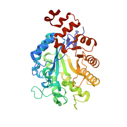
    Xenobiotic reductase A (XenA) from Pseudomonas putida 86 catalyzes the NAD(P)H-dependent reduction of various α,β-unsaturated carbonyl compounds and is a member of the old yellow enzyme family. The reaction of XenA follows a ping-pong mechanism, implying that its active site has to accommodate and correctly position the various substrates to be oxidized (NADH/NADPH) and to be reduced (different α,β-unsaturated carbonyl compounds) to enable formal hydride transfers between the substrate and the isoalloxazine ring. The active site of XenA is lined by two tyrosine (Tyr27, Tyr183) and two tryptophan (Trp302, Trp358) residues, which were proposed to contribute to substrate binding. We analyzed the individual contributions of the four residues, using site-directed mutagenesis, steady-state and transient kinetics, redox potentiometry and crystal structure analysis. The Y183F substitution decreases the affinity of XenA for NADPH and reduces the rate of the oxidative half-reaction by two to three orders of magnitude, the latter being in agreement with its function as a proton donor in the oxidative half-reaction. Upon reduction of the flavin, Trp302 swings into the active site of XenA (in-conformation) and decreases the extent of the substrate-binding pocket. Its exchange against alanine induces substrate inhibition at elevated NADPH concentrations, indicating that the in-conformation of Trp302 helps to disfavor the nonproductive NADPH binding in the reduced state of XenA. Our analysis shows that while the principal catalytic mechanism of XenA, for example, type of proton donor, is analogous to that of other members of the old yellow enzyme family, its strategy to correctly position and accommodate different substrates is unprecedented.


  • Organizational Affiliation

    AG Bioanorganische Chemie, Universität Bayreuth, Universitätsstrasse 30, 95447 Bayreuth, Germany.


Macromolecules
Find similar proteins by:  (by identity cutoff)  |  3D Structure
Entity ID: 1
MoleculeChains Sequence LengthOrganismDetailsImage
Xenobiotic reductase A363Pseudomonas putidaMutation(s): 0 
Gene Names: xenA
EC: 1.6.99.1
UniProt
Find proteins for Q3ZDM6 (Pseudomonas putida)
Explore Q3ZDM6 
Go to UniProtKB:  Q3ZDM6
Entity Groups  
Sequence Clusters30% Identity50% Identity70% Identity90% Identity95% Identity100% Identity
UniProt GroupQ3ZDM6
Sequence Annotations
Expand
  • Reference Sequence
Small Molecules
Ligands 3 Unique
IDChains Name / Formula / InChI Key2D Diagram3D Interactions
FMN
Query on FMN

Download Ideal Coordinates CCD File 
B [auth A]FLAVIN MONONUCLEOTIDE
C17 H21 N4 O9 P
FVTCRASFADXXNN-SCRDCRAPSA-N
SO4
Query on SO4

Download Ideal Coordinates CCD File 
C [auth A]
D [auth A]
E [auth A]
F [auth A]
G [auth A]
C [auth A],
D [auth A],
E [auth A],
F [auth A],
G [auth A],
H [auth A]
SULFATE ION
O4 S
QAOWNCQODCNURD-UHFFFAOYSA-L
BU3
Query on BU3

Download Ideal Coordinates CCD File 
I [auth A](R,R)-2,3-BUTANEDIOL
C4 H10 O2
OWBTYPJTUOEWEK-QWWZWVQMSA-N
Experimental Data & Validation

Experimental Data

  • Method: X-RAY DIFFRACTION
  • Resolution: 1.90 Å
  • R-Value Free: 0.234 
  • R-Value Work: 0.187 
  • R-Value Observed: 0.189 
  • Space Group: I 2 2 2
Unit Cell:
Length ( Å )Angle ( ˚ )
a = 58.347α = 90
b = 83.534β = 90
c = 156.33γ = 90
Software Package:
Software NamePurpose
HKL-2000data collection
PHENIXmodel building
PHENIXrefinement
XDSdata reduction
XSCALEdata scaling
PHENIXphasing

Structure Validation

View Full Validation Report


Ligand Structure Quality Assessment 


Entry History 

Deposition Data

Revision History  (Full details and data files)

  • Version 1.0: 2010-09-22
    Type: Initial release
  • Version 1.1: 2011-07-13
    Changes: Version format compliance
  • Version 1.2: 2014-03-05
    Changes: Database references
  • Version 1.3: 2023-11-01
    Changes: Data collection, Database references, Derived calculations, Refinement description