3MWY

Crystal structure of the chromodomain-ATPase portion of the yeast Chd1 chromatin remodeler


Experimental Data Snapshot

  • Method: X-RAY DIFFRACTION
  • Resolution: 3.70 Å
  • R-Value Free: 0.318 
  • R-Value Work: 0.262 
  • R-Value Observed: 0.265 

wwPDB Validation   3D Report Full Report


Ligand Structure Quality Assessment 


This is version 1.4 of the entry. See complete history


Literature

The chromodomains of the Chd1 chromatin remodeler regulate DNA access to the ATPase motor.

Hauk, G.McKnight, J.N.Nodelman, I.M.Bowman, G.D.

(2010) Mol Cell 39: 711-723

  • DOI: https://doi.org/10.1016/j.molcel.2010.08.012
  • Primary Citation of Related Structures:  
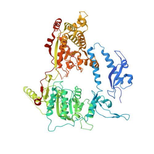
    3MWY

  • PubMed Abstract: 

    Chromatin remodelers are ATP-driven machines that assemble, slide, and remove nucleosomes from DNA, but how the ATPase motors of remodelers are regulated is poorly understood. Here we show that the double chromodomain unit of the Chd1 remodeler blocks DNA binding and activation of the ATPase motor in the absence of nucleosome substrates. The Chd1 crystal structure reveals that an acidic helix joining the chromodomains can pack against a DNA-binding surface of the ATPase motor. Disruption of the chromodomain-ATPase interface prevents discrimination between nucleosomes and naked DNA and reduces the reliance on the histone H4 tail for nucleosome sliding. We propose that the chromodomains allow Chd1 to distinguish between nucleosomes and naked DNA by physically gating access to the ATPase motor, and we hypothesize that related ATPase motors may employ a similar strategy to discriminate among DNA-containing substrates.


  • Organizational Affiliation

    TC Jenkins Department of Biophysics, Johns Hopkins University, Baltimore, MD 21218-2685, USA.


Macromolecules
Find similar proteins by:  (by identity cutoff)  |  3D Structure
Entity ID: 1
MoleculeChains Sequence LengthOrganismDetailsImage
Chromo domain-containing protein 1A [auth W]800Saccharomyces cerevisiaeMutation(s): 0 
Gene Names: CHD1SYGP-ORF4YER164W
EC: 3.6.1
UniProt
Find proteins for P32657 (Saccharomyces cerevisiae (strain ATCC 204508 / S288c))
Explore P32657 
Go to UniProtKB:  P32657
Entity Groups  
Sequence Clusters30% Identity50% Identity70% Identity90% Identity95% Identity100% Identity
UniProt GroupP32657
Sequence Annotations
Expand
  • Reference Sequence
Small Molecules
Ligands 1 Unique
IDChains Name / Formula / InChI Key2D Diagram3D Interactions
AGS
Query on AGS

Download Ideal Coordinates CCD File 
B [auth W]PHOSPHOTHIOPHOSPHORIC ACID-ADENYLATE ESTER
C10 H16 N5 O12 P3 S
NLTUCYMLOPLUHL-KQYNXXCUSA-N
Experimental Data & Validation

Experimental Data

  • Method: X-RAY DIFFRACTION
  • Resolution: 3.70 Å
  • R-Value Free: 0.318 
  • R-Value Work: 0.262 
  • R-Value Observed: 0.265 
  • Space Group: P 61 2 2
Unit Cell:
Length ( Å )Angle ( ˚ )
a = 94.333α = 90
b = 94.333β = 90
c = 450.088γ = 120
Software Package:
Software NamePurpose
REFMACrefinement
SOLVEphasing
SHARPphasing
PHENIXrefinement
CBASSdata collection
HKL-2000data reduction
SCALEPACKdata scaling

Structure Validation

View Full Validation Report


Ligand Structure Quality Assessment 


Entry History 

Deposition Data

Revision History  (Full details and data files)

  • Version 1.0: 2010-09-29
    Type: Initial release
  • Version 1.1: 2011-07-13
    Changes: Version format compliance
  • Version 1.2: 2013-01-30
    Changes: Non-polymer description
  • Version 1.3: 2019-07-17
    Changes: Data collection, Refinement description
  • Version 1.4: 2023-09-06
    Changes: Data collection, Database references, Derived calculations, Refinement description