3MQI

Human early B-cell factor 1 (EBF1) IPT/TIG domain


Experimental Data Snapshot

  • Method: X-RAY DIFFRACTION
  • Resolution: 2.30 Å
  • R-Value Free: 0.273 
  • R-Value Work: 0.233 
  • R-Value Observed: 0.235 

wwPDB Validation   3D Report Full Report


This is version 1.2 of the entry. See complete history


Literature

Structural Determination of Functional Domains in Early B-cell Factor (EBF) Family of Transcription Factors Reveals Similarities to Rel DNA-binding Proteins and a Novel Dimerization Motif.

Siponen, M.I.Wisniewska, M.Lehtio, L.Johansson, I.Svensson, L.Raszewski, G.Nilsson, L.Sigvardsson, M.Berglund, H.

(2010) J Biol Chem 285: 25875-25879

  • DOI: https://doi.org/10.1074/jbc.C110.150482
  • Primary Citation of Related Structures:  
    3LYR, 3MQI, 3MUJ, 3N50

  • PubMed Abstract: 

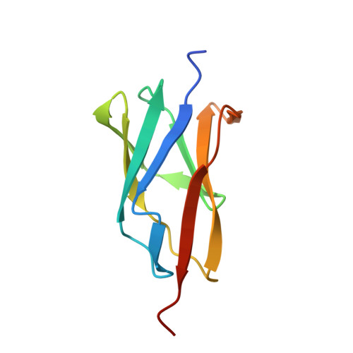
    The early B-cell factor (EBF) transcription factors are central regulators of development in several organs and tissues. This protein family shows low sequence similarity to other protein families, which is why structural information for the functional domains of these proteins is crucial to understand their biochemical features. We have used a modular approach to determine the crystal structures of the structured domains in the EBF family. The DNA binding domain reveals a striking resemblance to the DNA binding domains of the Rel homology superfamily of transcription factors but contains a unique zinc binding structure, termed zinc knuckle. Further the EBF proteins contain an IPT/TIG domain and an atypical helix-loop-helix domain with a novel type of dimerization motif. The data presented here provide insights into unique structural features of the EBF proteins and open possibilities for detailed molecular investigations of this important transcription factor family.


  • Organizational Affiliation

    Department of Medical Biochemistry and Biophysics, Karolinska Institutet, Stockholm, Sweden.


Macromolecules
Find similar proteins by:  (by identity cutoff)  |  3D Structure
Entity ID: 1
MoleculeChains Sequence LengthOrganismDetailsImage
Transcription factor COE1
A, B, C
96Homo sapiensMutation(s): 0 
Gene Names: COE1EBFEBF1EBF1A
UniProt & NIH Common Fund Data Resources
Find proteins for Q9UH73 (Homo sapiens)
Explore Q9UH73 
Go to UniProtKB:  Q9UH73
PHAROS:  Q9UH73
GTEx:  ENSG00000164330 
Entity Groups  
Sequence Clusters30% Identity50% Identity70% Identity90% Identity95% Identity100% Identity
UniProt GroupQ9UH73
Sequence Annotations
Expand
  • Reference Sequence
Experimental Data & Validation

Experimental Data

  • Method: X-RAY DIFFRACTION
  • Resolution: 2.30 Å
  • R-Value Free: 0.273 
  • R-Value Work: 0.233 
  • R-Value Observed: 0.235 
  • Space Group: C 1 2 1
Unit Cell:
Length ( Å )Angle ( ˚ )
a = 86.291α = 90
b = 57.073β = 93.17
c = 69.14γ = 90
Software Package:
Software NamePurpose
APEXdata collection
SOLVEphasing
REFMACrefinement
XDSdata reduction
XSCALEdata scaling

Structure Validation

View Full Validation Report


Entry History 

Revision History  (Full details and data files)

  • Version 1.0: 2010-05-26
    Type: Initial release
  • Version 1.1: 2011-07-13
    Changes: Version format compliance
  • Version 1.2: 2024-02-21
    Changes: Data collection, Database references, Derived calculations