3MPW

Structure of EUTM in 2-D protein membrane


Experimental Data Snapshot

  • Method: X-RAY DIFFRACTION
  • Resolution: 2.70 Å
  • R-Value Free: 0.336 
  • R-Value Work: 0.248 
  • R-Value Observed: 0.253 

wwPDB Validation   3D Report Full Report


This is version 1.2 of the entry. See complete history


Literature

Crystallographic insights into the pore structures and mechanisms of the EutL and EutM shell proteins of the ethanolamine-utilizing microcompartment of Escherichia coli.

Takenoya, M.Nikolakakis, K.Sagermann, M.

(2010) J Bacteriol 192: 6056-6063

  • DOI: https://doi.org/10.1128/JB.00652-10
  • Primary Citation of Related Structures:  
    3MPV, 3MPW, 3MPY

  • PubMed Abstract: 

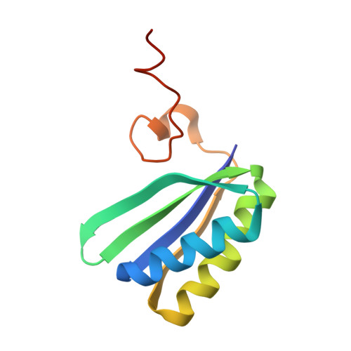
    The ethanolamine-utilizing bacterial microcompartment (Eut-BMC) of Escherichia coli is a polyhedral organelle that harbors specific enzymes for the catabolic degradation of ethanolamine. The compartment is composed of a proteinaceous shell structure that maintains a highly specialized environment for the biochemical reactions inside. Recent structural investigations have revealed hexagonal assemblies of shell proteins that form a tightly packed two-dimensional lattice that is likely to function as a selectively permeable protein membrane, wherein small channels are thought to permit controlled exchange of specific solutes. Here, we show with two nonisomorphous crystal structures that EutM also forms a two-dimensional protein membrane. As its architecture is highly similar to the membrane structure of EutL, it is likely that the structure represents a physiologically relevant form. Thus far, of all Eut proteins, only EutM and EutL have been shown to form such proteinaceous membranes. Despite their similar architectures, however, both proteins exhibit dramatically different pore structures. In contrast to EutL, the pore of EutM appears to be positively charged, indicating specificity for different solutes. Furthermore, we also show that the central pore structure of the EutL shell protein can be triggered to open specifically upon exposure to zinc ions, suggesting a specific gating mechanism.


  • Organizational Affiliation

    Department of Biotechnology and Life Science, Tokyo University of Agriculture & Technology, Tokyo, Japan.


Macromolecules
Find similar proteins by:  (by identity cutoff)  |  3D Structure
Entity ID: 1
MoleculeChains Sequence LengthOrganismDetailsImage
Ethanolamine utilization protein eutM103Escherichia coli K-12Mutation(s): 0 
Gene Names: b2457cchAeutMJW2441yffZ
UniProt
Find proteins for P0ABF4 (Escherichia coli (strain K12))
Explore P0ABF4 
Go to UniProtKB:  P0ABF4
Entity Groups  
Sequence Clusters30% Identity50% Identity70% Identity90% Identity95% Identity100% Identity
UniProt GroupP0ABF4
Sequence Annotations
Expand
  • Reference Sequence
Small Molecules
Ligands 1 Unique
IDChains Name / Formula / InChI Key2D Diagram3D Interactions
PO4
Query on PO4

Download Ideal Coordinates CCD File 
M [auth G]
N [auth H]
O [auth I]
P [auth I]
Q [auth J]
M [auth G],
N [auth H],
O [auth I],
P [auth I],
Q [auth J],
R [auth K],
S [auth L],
T [auth A],
U [auth B],
V [auth C],
W [auth C],
X [auth D],
Y [auth E],
Z [auth F]
PHOSPHATE ION
O4 P
NBIIXXVUZAFLBC-UHFFFAOYSA-K
Experimental Data & Validation

Experimental Data

  • Method: X-RAY DIFFRACTION
  • Resolution: 2.70 Å
  • R-Value Free: 0.336 
  • R-Value Work: 0.248 
  • R-Value Observed: 0.253 
  • Space Group: P 1 21 1
Unit Cell:
Length ( Å )Angle ( ˚ )
a = 69.949α = 90
b = 149.069β = 120.01
c = 70.089γ = 90
Software Package:
Software NamePurpose
REFMACrefinement

Structure Validation

View Full Validation Report


Entry History 

Deposition Data

Revision History  (Full details and data files)

  • Version 1.0: 2011-05-11
    Type: Initial release
  • Version 1.1: 2011-07-13
    Changes: Version format compliance
  • Version 1.2: 2023-09-06
    Changes: Data collection, Database references, Derived calculations, Refinement description