3MPP

Botulinum Neurotoxin Type G Receptor Binding Domain


Experimental Data Snapshot

  • Method: X-RAY DIFFRACTION
  • Resolution: 1.98 Å
  • R-Value Free: 0.221 
  • R-Value Work: 0.175 
  • R-Value Observed: 0.177 

wwPDB Validation   3D Report Full Report


This is version 1.3 of the entry. See complete history


Literature

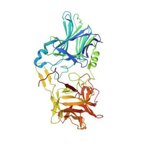
Structural analysis of botulinum neurotoxin type G receptor binding .

Schmitt, J.Karalewitz, A.Benefield, D.A.Mushrush, D.J.Pruitt, R.N.Spiller, B.W.Barbieri, J.T.Lacy, D.B.

(2010) Biochemistry 49: 5200-5205

  • DOI: https://doi.org/10.1021/bi100412v
  • Primary Citation of Related Structures:  
    3MPP

  • PubMed Abstract: 

    Botulinum neurotoxin (BoNT) binds peripheral neurons at the neuromuscular junction through a dual-receptor mechanism that includes interactions with ganglioside and protein receptors. The receptor identities vary depending on BoNT serotype (A-G). BoNT/B and BoNT/G bind the luminal domains of synaptotagmin I and II, homologous synaptic vesicle proteins. We observe conditions under which BoNT/B binds both Syt isoforms, but BoNT/G binds only SytI. Both serotypes bind ganglioside G(T1b). The BoNT/G receptor-binding domain crystal structure provides a context for examining these binding interactions and a platform for understanding the physiological relevance of different Syt receptor isoforms in vivo.


  • Organizational Affiliation

    Department of Microbiology and Immunology, Vanderbilt University School of Medicine, Nashville, Tennessee 37232, USA.


Macromolecules
Find similar proteins by:  (by identity cutoff)  |  3D Structure
Entity ID: 1
MoleculeChains Sequence LengthOrganismDetailsImage
Botulinum neurotoxin type GA [auth G]433Clostridium botulinumMutation(s): 4 
Gene Names: botG
EC: 3.4.24.69
UniProt
Find proteins for Q60393 (Clostridium botulinum)
Explore Q60393 
Go to UniProtKB:  Q60393
Entity Groups  
Sequence Clusters30% Identity50% Identity70% Identity90% Identity95% Identity100% Identity
UniProt GroupQ60393
Sequence Annotations
Expand
  • Reference Sequence
Experimental Data & Validation

Experimental Data

  • Method: X-RAY DIFFRACTION
  • Resolution: 1.98 Å
  • R-Value Free: 0.221 
  • R-Value Work: 0.175 
  • R-Value Observed: 0.177 
  • Space Group: P 21 21 21
Unit Cell:
Length ( Å )Angle ( ˚ )
a = 57.63α = 90
b = 90.161β = 90
c = 91.894γ = 90
Software Package:
Software NamePurpose
HKL-2000data collection
MOLREPphasing
PHENIXrefinement
HKL-2000data reduction
HKL-2000data scaling

Structure Validation

View Full Validation Report


Entry History 

Deposition Data

Revision History  (Full details and data files)

  • Version 1.0: 2010-06-16
    Type: Initial release
  • Version 1.1: 2011-07-13
    Changes: Version format compliance
  • Version 1.2: 2021-10-06
    Changes: Database references
  • Version 1.3: 2023-09-06
    Changes: Data collection, Refinement description