The Receptor-Binding Domain of Influenza Virus Hemagglutinin Produced in Escherichia coli Folds into Its Native, Immunogenic Structure.
Dubois, R.M., Aguilar-Yanez, J.M., Mendoza-Ochoa, G.I., Oropeza-Almazan, Y., Schultz-Cherry, S., Alvarez, M.M., White, S.W., Russell, C.J.(2011) J Virol 85: 865-872
- PubMed: 21068239
- DOI: https://doi.org/10.1128/JVI.01412-10
- Primary Citation of Related Structures:
3MLH - PubMed Abstract:
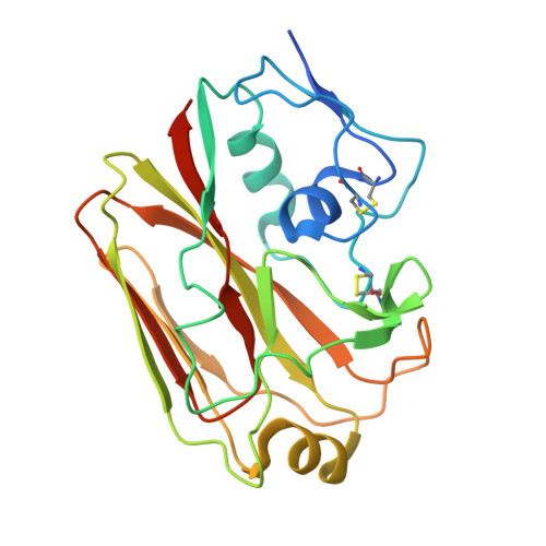
The hemagglutinin (HA) surface glycoprotein promotes influenza virus entry and is the key protective antigen in natural immunity and vaccines. The HA protein is a trimeric envelope glycoprotein consisting of a globular receptor-binding domain (HA-RBD) that is inserted into a membrane fusion-mediating stalk domain. Similar to other class I viral fusion proteins, the fusogenic stalk domain spontaneously refolds into its postfusion conformation when expressed in isolation, consistent with this domain being trapped in a metastable conformation. Using X-ray crystallography, we show that the influenza virus HA-RBD refolds spontaneously into its native, immunogenic structure even when expressed in an unglycosylated form in Escherichia coli. In the 2.10-Å structure of the HA-RBD, the receptor-binding pocket is intact and its conformational epitopes are preserved. Recombinant HA-RBD is immunogenic and protective in ferrets, and the protein also binds with specificity to sera from influenza virus-infected humans. Overall, the data provide a structural basis for the rapid production of influenza vaccines in E. coli. From an evolutionary standpoint, the ability of the HA-RBD to refold spontaneously into its native conformation suggests that influenza virus acquired this domain as an insertion into an ancestral membrane-fusion domain. The insertion of independently folding domains into fusogenic stalk domains may be a common feature of class I viral fusion proteins.
Organizational Affiliation:
Department of Structural Biology, St. Jude Children's Research Hospital, 262 Danny Thomas Place, Memphis, TN 38105, USA.