3MEH

Crystal structure of Staphylococcal nuclease variant Delta+PHS I92A at cryogenic temperature


Experimental Data Snapshot

  • Method: X-RAY DIFFRACTION
  • Resolution: 1.50 Å
  • R-Value Free: 0.234 
  • R-Value Work: 0.186 
  • R-Value Observed: 0.191 

wwPDB Validation   3D Report Full Report


Ligand Structure Quality Assessment 


This is version 1.7 of the entry. See complete history


Literature

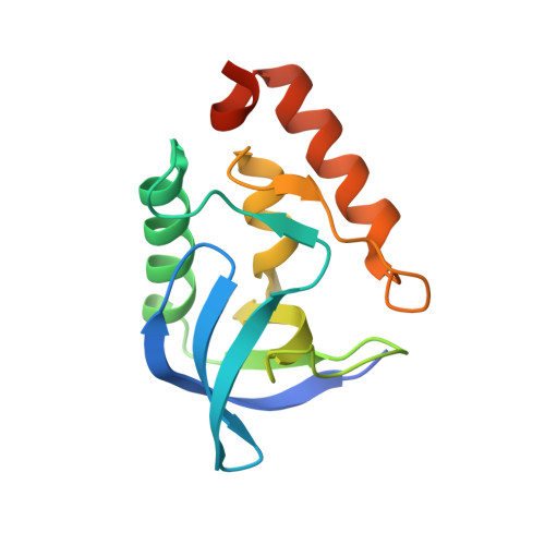
Cavities determine the pressure unfolding of proteins.

Roche, J.Caro, J.A.Norberto, D.R.Barthe, P.Roumestand, C.Schlessman, J.L.Garcia, A.E.Garcia-Moreno E, B.Royer, C.A.

(2012) Proc Natl Acad Sci U S A 109: 6945-6950

  • DOI: https://doi.org/10.1073/pnas.1200915109
  • Primary Citation of Related Structures:  
    3MEH, 3MHB, 3MVV, 3MXP, 3MZ5, 3NK9, 3NP8, 3NQT, 3NXW, 3OSO, 3PMF, 3R3O

  • PubMed Abstract: 

    It has been known for nearly 100 years that pressure unfolds proteins, yet the physical basis of this effect is not understood. Unfolding by pressure implies that the molar volume of the unfolded state of a protein is smaller than that of the folded state. This decrease in volume has been proposed to arise from differences between the density of bulk water and water associated with the protein, from pressure-dependent changes in the structure of bulk water, from the loss of internal cavities in the folded states of proteins, or from some combination of these three factors. Here, using 10 cavity-containing variants of staphylococcal nuclease, we demonstrate that pressure unfolds proteins primarily as a result of cavities that are present in the folded state and absent in the unfolded one. High-pressure NMR spectroscopy and simulations constrained by the NMR data were used to describe structural and energetic details of the folding landscape of staphylococcal nuclease that are usually inaccessible with existing experimental approaches using harsher denaturants. Besides solving a 100-year-old conundrum concerning the detailed structural origins of pressure unfolding of proteins, these studies illustrate the promise of pressure perturbation as a unique tool for examining the roles of packing, conformational fluctuations, and water penetration as determinants of solution properties of proteins, and for detecting folding intermediates and other structural details of protein-folding landscapes that are invisible to standard experimental approaches.


  • Organizational Affiliation

    Centre de Biochimie Structurale, Institut National pour la Santé et la Recherche Médicale U554, Centre National pour la Recherche Scientifique Unité Mixte de Recherche 5048, Université Montpellier 1&2, Montpellier, France.


Macromolecules
Find similar proteins by:  (by identity cutoff)  |  3D Structure
Entity ID: 1
MoleculeChains Sequence LengthOrganismDetailsImage
Thermonuclease143Staphylococcus aureusMutation(s): 6 
EC: 3.1.31.1
UniProt
Find proteins for P00644 (Staphylococcus aureus)
Explore P00644 
Go to UniProtKB:  P00644
Entity Groups  
Sequence Clusters30% Identity50% Identity70% Identity90% Identity95% Identity100% Identity
UniProt GroupP00644
Sequence Annotations
Expand
  • Reference Sequence
Small Molecules
Ligands 2 Unique
IDChains Name / Formula / InChI Key2D Diagram3D Interactions
THP
Query on THP

Download Ideal Coordinates CCD File 
C [auth A]THYMIDINE-3',5'-DIPHOSPHATE
C10 H16 N2 O11 P2
CSNCBOPUCJOHLS-XLPZGREQSA-N
CA
Query on CA

Download Ideal Coordinates CCD File 
B [auth A]CALCIUM ION
Ca
BHPQYMZQTOCNFJ-UHFFFAOYSA-N
Experimental Data & Validation

Experimental Data

  • Method: X-RAY DIFFRACTION
  • Resolution: 1.50 Å
  • R-Value Free: 0.234 
  • R-Value Work: 0.186 
  • R-Value Observed: 0.191 
  • Space Group: P 1 21 1
Unit Cell:
Length ( Å )Angle ( ˚ )
a = 31.09α = 90
b = 60.137β = 93.57
c = 38.194γ = 90
Software Package:
Software NamePurpose
DENZOdata reduction
SCALEPACKdata scaling
PHASERphasing
REFMACrefinement
PDB_EXTRACTdata extraction
CBASSdata collection
HKL-2000data reduction

Structure Validation

View Full Validation Report


Ligand Structure Quality Assessment 


Entry History 

Deposition Data

Revision History  (Full details and data files)

  • Version 1.0: 2011-03-16
    Type: Initial release
  • Version 1.1: 2011-07-13
    Changes: Version format compliance
  • Version 1.2: 2011-09-07
    Changes: Atomic model
  • Version 1.3: 2012-05-23
    Changes: Database references
  • Version 1.4: 2014-06-04
    Changes: Atomic model
  • Version 1.5: 2017-11-08
    Changes: Refinement description
  • Version 1.6: 2021-10-06
    Changes: Database references, Derived calculations
  • Version 1.7: 2023-09-06
    Changes: Data collection, Refinement description