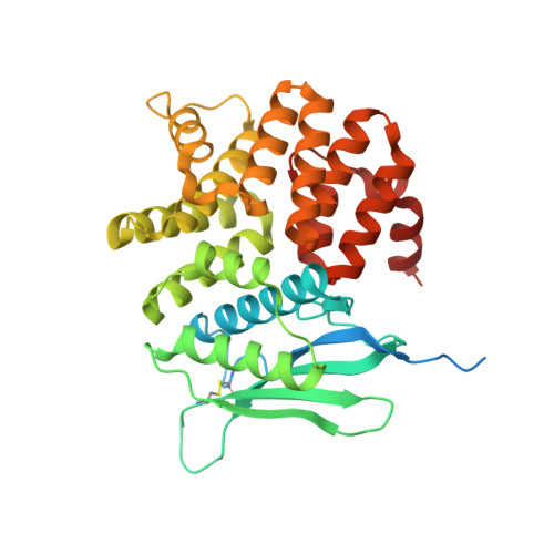
Crystal structure of the sensory domain of Escherichia coli CadC, a member of the ToxR-like protein family.
Eichinger, A., Haneburger, I., Koller, C., Jung, K., Skerra, A.(2011) Protein Sci 20: 656-669
- PubMed: 21308846
- DOI: https://doi.org/10.1002/pro.594
- Primary Citation of Related Structures:
3LY7, 3LY8, 3LY9, 3LYA - PubMed Abstract:
The membrane-integral transcriptional activator CadC comprises sensory and transcriptional regulatory functions within one polypeptide chain. Its C-terminal periplasmic domain, CadC(pd), is responsible for sensing of environmental pH as well as for binding of the feedback inhibitor cadaverine. Here we describe the crystal structure of CadC(pd) (residues 188-512) solved at a resolution of 1.8 Å via multiple wavelength anomalous dispersion (MAD) using a ReCl(6)(2-) derivative. CadC(pd) reveals a novel fold comprising two subdomains: an N-terminal subdomain dominated by a β-sheet in contact with three α-helices and a C-terminal subdomain formed by an eleven-membered α-helical bundle, which is oriented almost perpendicular to the helices in the first subdomain. Further to the native protein, crystal structures were also solved for its variants D471N and D471E, which show functionally different behavior in pH sensing. Interestingly, in the heavy metal derivative of CadC(pd) used for MAD phasing a ReCl(6)(2-) ion was found in a cavity located between the two subdomains. Amino acid side chains that coordinate this complex ion are conserved in CadC homologues from various bacterial species, suggesting a function of the cavity in the binding of cadaverine, which was supported by docking studies. Notably, CadC(pd) forms a homo-dimer in solution, which can be explained by an extended, albeit rather polar interface between two symmetry-related monomers in the crystal structure. The occurrence of several acidic residues in this region suggests protonation-dependent changes in the mode of dimerization, which could eventually trigger transcriptional activation by CadC in the bacterial cytoplasm.
Organizational Affiliation:
Munich Center for Integrated Protein Science and Lehrstuhl für Biologische Chemie, Technische Universität München, Freising-Weihenstephan, Germany.