3LTR

5-OMe-dU containing DNA 8mer


Experimental Data Snapshot

  • Method: X-RAY DIFFRACTION
  • Resolution: 1.30 Å
  • R-Value Free: 0.222 
  • R-Value Work: 0.202 
  • R-Value Observed: 0.203 

wwPDB Validation   3D Report Full Report


This is version 1.4 of the entry. See complete history


Literature

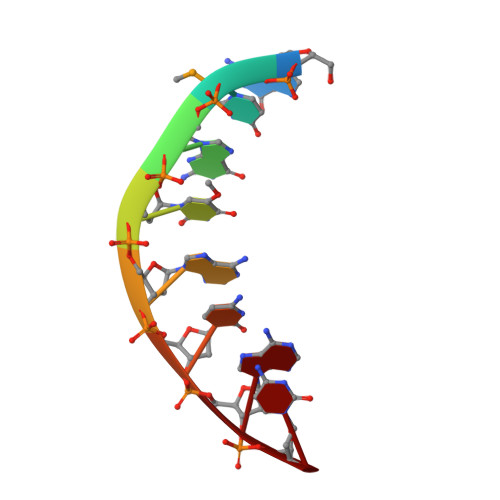
Hydrogen bond formation between the naturally modified nucleobase and phosphate backbone.

Sheng, J.Zhang, W.Hassan, A.E.Gan, J.Soares, A.S.Geng, S.Ren, Y.Huang, Z.

(2012) Nucleic Acids Res 40: 8111-8118

  • DOI: https://doi.org/10.1093/nar/gks426
  • Primary Citation of Related Structures:  
    3IKI, 3LTR

  • PubMed Abstract: 

    Natural RNAs, especially tRNAs, are extensively modified to tailor structure and function diversities. Uracil is the most modified nucleobase among all natural nucleobases. Interestingly, >76% of uracil modifications are located on its 5-position. We have investigated the natural 5-methoxy (5-O-CH(3)) modification of uracil in the context of A-form oligonucleotide duplex. Our X-ray crystal structure indicates first a H-bond formation between the uracil 5-O-CH(3) and its 5'-phosphate. This novel H-bond is not observed when the oxygen of 5-O-CH(3) is replaced with a larger atom (selenium or sulfur). The 5-O-CH(3) modification does not cause significant structure and stability alterations. Moreover, our computational study is consistent with the experimental observation. The investigation on the uracil 5-position demonstrates the importance of this RNA modification at the atomic level. Our finding suggests a general interaction between the nucleobase and backbone and reveals a plausible function of the tRNA 5-O-CH(3) modification, which might potentially rigidify the local conformation and facilitates translation.


  • Organizational Affiliation

    Department of Chemistry, Georgia State University, Atlanta, GA 30303, USA.


Macromolecules

Find similar nucleic acids by:  Sequence   |   3D Structure  

Entity ID: 1
MoleculeChains LengthOrganismImage
5'-D(*GP*(UMS)P*GP*(T5O)P*AP*CP*AP*C)-3'8N/A
Sequence Annotations
Expand
  • Reference Sequence
Small Molecules
Ligands 1 Unique
IDChains Name / Formula / InChI Key2D Diagram3D Interactions
MG
Query on MG

Download Ideal Coordinates CCD File 
B [auth A]MAGNESIUM ION
Mg
JLVVSXFLKOJNIY-UHFFFAOYSA-N
Experimental Data & Validation

Experimental Data

  • Method: X-RAY DIFFRACTION
  • Resolution: 1.30 Å
  • R-Value Free: 0.222 
  • R-Value Work: 0.202 
  • R-Value Observed: 0.203 
  • Space Group: P 43 21 2
Unit Cell:
Length ( Å )Angle ( ˚ )
a = 42.992α = 90
b = 42.992β = 90
c = 23.822γ = 90
Software Package:
Software NamePurpose
CBASSdata collection
PHASESphasing
REFMACrefinement
HKL-2000data reduction
HKL-2000data scaling

Structure Validation

View Full Validation Report


Entry History 

Deposition Data

Revision History  (Full details and data files)

  • Version 1.0: 2010-03-09
    Type: Initial release
  • Version 1.1: 2011-07-13
    Changes: Advisory, Version format compliance
  • Version 1.2: 2012-06-13
    Changes: Database references
  • Version 1.3: 2012-09-26
    Changes: Database references
  • Version 1.4: 2023-09-06
    Changes: Data collection, Database references, Derived calculations, Refinement description