3LPZ

Crystal structure of C. therm. Get4


Experimental Data Snapshot

  • Method: X-RAY DIFFRACTION
  • Resolution: 1.98 Å
  • R-Value Free: 0.222 
  • R-Value Work: 0.166 
  • R-Value Observed: 0.169 

wwPDB Validation   3D Report Full Report


This is version 2.1 of the entry. See complete history


Literature

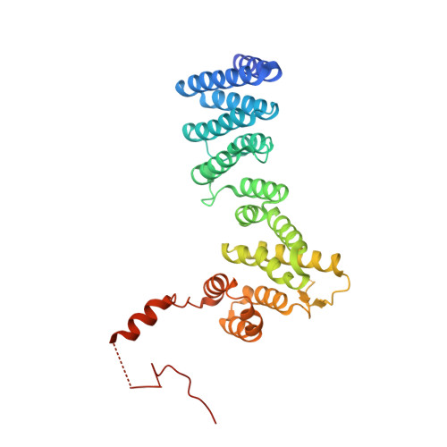
The structure of Get4 reveals an alpha-solenoid fold adapted for multiple interactions in tail-anchored protein biogenesis.

Bozkurt, G.Wild, K.Amlacher, S.Hurt, E.Dobberstein, B.Sinning, I.

(2010) FEBS Lett 584: 1509-1514

  • DOI: https://doi.org/10.1016/j.febslet.2010.02.070
  • Primary Citation of Related Structures:  
    3LPZ

  • PubMed Abstract: 

    Tail-anchored proteins play important roles in protein translocation, membrane fusion and apoptosis. They are targeted to the endoplasmic reticulum membrane via the guided-entry of tail-anchored proteins (Get) pathway. We present the 2A crystal structure of Get4 which participates in early steps of the Get pathway. The structure shows an alpha-solenoid fold with particular deviations from the regular pairwise arrangement of alpha-helices. A conserved hydrophobic groove accommodates the flexible C-terminal region in trans. The structural organization of the Get4 helical hairpin motifs provides a scaffold for protein-protein interactions in the Get pathway.


  • Organizational Affiliation

    Heidelberg University Biochemistry Center (BZH), Heidelberg, Germany.


Macromolecules
Find similar proteins by:  (by identity cutoff)  |  3D Structure
Entity ID: 1
MoleculeChains Sequence LengthOrganismDetailsImage
Uncharacterized protein336Thermochaetoides thermophila DSM 1495Mutation(s): 0 
Gene Names: CTHT_0028730
UniProt
Find proteins for G0S7T3 (Chaetomium thermophilum (strain DSM 1495 / CBS 144.50 / IMI 039719))
Explore G0S7T3 
Go to UniProtKB:  G0S7T3
Entity Groups  
Sequence Clusters30% Identity50% Identity70% Identity90% Identity95% Identity100% Identity
UniProt GroupG0S7T3
Sequence Annotations
Expand
  • Reference Sequence
Experimental Data & Validation

Experimental Data

  • Method: X-RAY DIFFRACTION
  • Resolution: 1.98 Å
  • R-Value Free: 0.222 
  • R-Value Work: 0.166 
  • R-Value Observed: 0.169 
  • Space Group: P 21 21 21
Unit Cell:
Length ( Å )Angle ( ˚ )
a = 54.92α = 90
b = 79.68β = 90
c = 94.71γ = 90
Software Package:
Software NamePurpose
REFMACrefinement
PDB_EXTRACTdata extraction
DNAdata collection
MOSFLMdata reduction
SCALAdata scaling
PHENIXphasing

Structure Validation

View Full Validation Report


Entry History 

Deposition Data

Revision History  (Full details and data files)

  • Version 1.0: 2010-03-16
    Type: Initial release
  • Version 1.1: 2011-07-13
    Changes: Version format compliance
  • Version 2.0: 2019-11-06
    Changes: Advisory, Atomic model, Data collection, Database references, Source and taxonomy, Structure summary
  • Version 2.1: 2024-02-21
    Changes: Data collection, Database references