3LAW

Structure of GTP-bound L129F mutant Rab7


Experimental Data Snapshot

  • Method: X-RAY DIFFRACTION
  • Resolution: 2.80 Å
  • R-Value Free: 0.273 
  • R-Value Work: 0.220 
  • R-Value Observed: 0.237 

wwPDB Validation   3D Report Full Report


Ligand Structure Quality Assessment 


This is version 1.3 of the entry. See complete history


Literature

Disease mutations in Rab7 result in unregulated nucleotide exchange and inappropriate activation.

McCray, B.A.Skordalakes, E.Taylor, J.P.

(2010) Hum Mol Genet 19: 1033-1047

  • DOI: https://doi.org/10.1093/hmg/ddp567
  • Primary Citation of Related Structures:  
    3LAW

  • PubMed Abstract: 

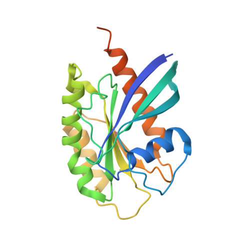
    Rab GTPases are molecular switches that orchestrate vesicular trafficking, maturation and fusion by cycling between an active, GTP-bound form, and an inactive, GDP-bound form. The activity cycle is coupled to GTP hydrolysis and is tightly controlled by regulatory proteins. Missense mutations of the GTPase Rab7 cause a dominantly inherited axonal degeneration known as Charcot-Marie-Tooth type 2B through an unknown mechanism. We present the 2.8 A crystal structure of GTP-bound L129F mutant Rab7 which reveals normal conformations of the effector binding regions and catalytic site, but an alteration to the nucleotide binding pocket that is predicted to alter GTP binding. Through extensive biochemical analysis, we demonstrate that disease-associated mutations in Rab7 do not lead to an intrinsic GTPase defect, but permit unregulated nucleotide exchange leading to both excessive activation and hydrolysis-independent inactivation. Consistent with augmented activity, mutant Rab7 shows significantly enhanced interaction with a subset of effector proteins. In addition, dynamic imaging demonstrates that mutant Rab7 is abnormally retained on target membranes. However, we show that the increased activation of mutant Rab7 is counterbalanced by unregulated, GTP hydrolysis-independent membrane cycling. Notably, disease mutations are able to rescue the membrane cycling of a GTPase-deficient mutant. Thus, we demonstrate that disease mutations uncouple Rab7 from the spatial and temporal control normally imposed by regulatory proteins and cause disease not by a gain of novel toxic function, but by misregulation of native Rab7 activity.


  • Organizational Affiliation

    Department of Developmental Neurobiology, St Jude Children's Research Hospital, Memphis, TN 38105-3678, USA.


Macromolecules
Find similar proteins by:  (by identity cutoff)  |  3D Structure
Entity ID: 1
MoleculeChains Sequence LengthOrganismDetailsImage
Ras-related protein Rab-7a
A, B, C, D, E
207Homo sapiensMutation(s): 1 
Gene Names: RAB7ARAB7
UniProt & NIH Common Fund Data Resources
Find proteins for P51149 (Homo sapiens)
Explore P51149 
Go to UniProtKB:  P51149
PHAROS:  P51149
GTEx:  ENSG00000075785 
Entity Groups  
Sequence Clusters30% Identity50% Identity70% Identity90% Identity95% Identity100% Identity
UniProt GroupP51149
Sequence Annotations
Expand
  • Reference Sequence
Small Molecules
Ligands 2 Unique
IDChains Name / Formula / InChI Key2D Diagram3D Interactions
GNP
Query on GNP

Download Ideal Coordinates CCD File 
G [auth A],
I [auth B],
K [auth C],
M [auth D],
O [auth E]
PHOSPHOAMINOPHOSPHONIC ACID-GUANYLATE ESTER
C10 H17 N6 O13 P3
UQABYHGXWYXDTK-UUOKFMHZSA-N
MG
Query on MG

Download Ideal Coordinates CCD File 
F [auth A],
H [auth B],
J [auth C],
L [auth D],
N [auth E]
MAGNESIUM ION
Mg
JLVVSXFLKOJNIY-UHFFFAOYSA-N
Experimental Data & Validation

Experimental Data

  • Method: X-RAY DIFFRACTION
  • Resolution: 2.80 Å
  • R-Value Free: 0.273 
  • R-Value Work: 0.220 
  • R-Value Observed: 0.237 
  • Space Group: P 1
Unit Cell:
Length ( Å )Angle ( ˚ )
a = 35.554α = 71.24
b = 89.335β = 85.97
c = 89.378γ = 85.91
Software Package:
Software NamePurpose
HKL-2000data collection
PHASERphasing
REFMACrefinement
HKL-2000data reduction
HKL-2000data scaling

Structure Validation

View Full Validation Report


Ligand Structure Quality Assessment 


Entry History 

Deposition Data

Revision History  (Full details and data files)

  • Version 1.0: 2010-01-26
    Type: Initial release
  • Version 1.1: 2011-07-13
    Changes: Advisory, Version format compliance
  • Version 1.2: 2021-10-13
    Changes: Database references, Derived calculations
  • Version 1.3: 2024-02-21
    Changes: Data collection