3L5T

Crystal structure of macrophage migration inhibitory factor (MIF) with thiophenepiperazinylquinolinone inhibitor at 1.86A resolution


Experimental Data Snapshot

  • Method: X-RAY DIFFRACTION
  • Resolution: 1.86 Å
  • R-Value Free: 0.214 
  • R-Value Work: 0.181 
  • R-Value Observed: 0.182 

wwPDB Validation   3D Report Full Report


Ligand Structure Quality Assessment 


This is version 1.3 of the entry. See complete history


Literature

Fragment screening of inhibitors for MIF tautomerase reveals a cryptic surface binding site.

McLean, L.R.Zhang, Y.Li, H.Choi, Y.M.Han, Z.Vaz, R.J.Li, Y.

(2010) Bioorg Med Chem Lett 20: 1821-1824

  • DOI: https://doi.org/10.1016/j.bmcl.2010.02.009
  • Primary Citation of Related Structures:  
    3L5P, 3L5R, 3L5S, 3L5T, 3L5U, 3L5V

  • PubMed Abstract: 

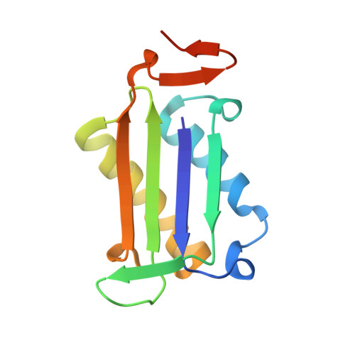
    In the course of a fragment screening campaign by in silico docking followed by X-ray crystallography, a novel binding site for migration inhibitory factor (MIF) inhibitors was demonstrated. The site is formed by rotation of the side-chain of Tyr-36 to reveal a surface binding site in MIF that is hydrophobic and surrounded by aromatic side-chain residues. The crystal structures of two small inhibitors that bind to this site and of a quinolinone inhibitor, that spans the canonical deep pocket near Pro-1 and the new surface binding site, have been solved. These results suggest new opportunities for structure-based design of MIF inhibitors.


  • Organizational Affiliation

    Discovery Research, Sanofi-aventis, Bridgewater, NJ 08807, USA. Larry.Mclean@sanofi-aventis.com


Macromolecules
Find similar proteins by:  (by identity cutoff)  |  3D Structure
Entity ID: 1
MoleculeChains Sequence LengthOrganismDetailsImage
Macrophage migration inhibitory factor
A, B, C
122Homo sapiensMutation(s): 0 
Gene Names: GLIFMIFMMIF
EC: 5.3.2.1 (PDB Primary Data), 5.3.3.12 (PDB Primary Data)
UniProt & NIH Common Fund Data Resources
Find proteins for P14174 (Homo sapiens)
Explore P14174 
Go to UniProtKB:  P14174
PHAROS:  P14174
GTEx:  ENSG00000240972 
Entity Groups  
Sequence Clusters30% Identity50% Identity70% Identity90% Identity95% Identity100% Identity
UniProt GroupP14174
Sequence Annotations
Expand
  • Reference Sequence
Small Molecules
Ligands 2 Unique
IDChains Name / Formula / InChI Key2D Diagram3D Interactions
956
Query on 956

Download Ideal Coordinates CCD File 
D [auth A]1-methyl-2-oxo-4-[4-(thiophen-2-ylcarbonyl)piperazin-1-yl]-1,2-dihydroquinoline-3-carbonitrile
C20 H18 N4 O2 S
GGGJOEPOPVIHEK-UHFFFAOYSA-N
SO4
Query on SO4

Download Ideal Coordinates CCD File 
E [auth B],
F [auth C]
SULFATE ION
O4 S
QAOWNCQODCNURD-UHFFFAOYSA-L
Experimental Data & Validation

Experimental Data

  • Method: X-RAY DIFFRACTION
  • Resolution: 1.86 Å
  • R-Value Free: 0.214 
  • R-Value Work: 0.181 
  • R-Value Observed: 0.182 
  • Space Group: P 21 21 21
Unit Cell:
Length ( Å )Angle ( ˚ )
a = 67.77α = 90
b = 67.661β = 90
c = 88.999γ = 90
Software Package:
Software NamePurpose
DENZOdata reduction
SCALEPACKdata scaling
MOLREPphasing
CNSrefinement
PDB_EXTRACTdata extraction
CrystalCleardata collection
CNXrefinement

Structure Validation

View Full Validation Report


Ligand Structure Quality Assessment 


Entry History 

Deposition Data

Revision History  (Full details and data files)

  • Version 1.0: 2010-03-09
    Type: Initial release
  • Version 1.1: 2011-07-13
    Changes: Version format compliance
  • Version 1.2: 2017-11-01
    Changes: Refinement description
  • Version 1.3: 2023-09-06
    Changes: Data collection, Database references, Derived calculations, Refinement description