3KX8

Structural basis of the activity and substrate specificity of the fluoroacetyl-CoA thioesterase FlK


Experimental Data Snapshot

  • Method: X-RAY DIFFRACTION
  • Resolution: 2.35 Å
  • R-Value Free: 0.243 
  • R-Value Work: 0.190 
  • R-Value Observed: 0.192 

wwPDB Validation   3D Report Full Report


This is version 1.4 of the entry. See complete history


Literature

Structural basis for the activity and substrate specificity of fluoroacetyl-CoA thioesterase FlK.

Dias, M.V.Huang, F.Chirgadze, D.Y.Tosin, M.Spiteller, D.Dry, E.F.Leadlay, P.F.Spencer, J.B.Blundell, T.L.

(2010) J Biol Chem 285: 22495-22504

  • DOI: https://doi.org/10.1074/jbc.M110.107177
  • Primary Citation of Related Structures:  
    3KUV, 3KUW, 3KV7, 3KV8, 3KVI, 3KVU, 3KVZ, 3KW1, 3KX7, 3KX8

  • PubMed Abstract: 

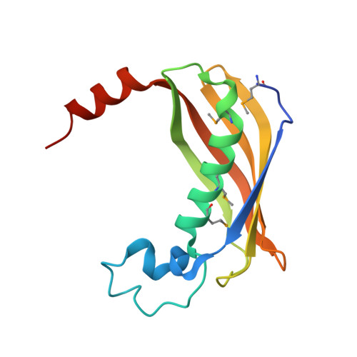
    The thioesterase FlK from the fluoroacetate-producing Streptomyces cattleya catalyzes the hydrolysis of fluoroacetyl-coenzyme A. This provides an effective self-defense mechanism, preventing any fluoroacetyl-coenzyme A formed from being further metabolized to 4-hydroxy-trans-aconitate, a lethal inhibitor of the tricarboxylic acid cycle. Remarkably, FlK does not accept acetyl-coenzyme A as a substrate. Crystal structure analysis shows that FlK forms a dimer, in which each subunit adopts a hot dog fold as observed for type II thioesterases. Unlike other type II thioesterases, which invariably utilize either an aspartate or a glutamate as catalytic base, we show by site-directed mutagenesis and crystallography that FlK employs a catalytic triad composed of Thr(42), His(76), and a water molecule, analogous to the Ser/Cys-His-acid triad of type I thioesterases. Structural comparison of FlK complexed with various substrate analogues suggests that the interaction between the fluorine of the substrate and the side chain of Arg(120) located opposite to the catalytic triad is essential for correct coordination of the substrate at the active site and therefore accounts for the substrate specificity.


  • Organizational Affiliation

    Department of Biochemistry, University of Cambridge, Lensfield Road, Cambridge CB2 1EW, United Kingdom.


Macromolecules
Find similar proteins by:  (by identity cutoff)  |  3D Structure
Entity ID: 1
MoleculeChains Sequence LengthOrganismDetailsImage
fluoroacetyl-CoA thioesterase
A, B, C, D, E
A, B, C, D, E, F, G, H
139Streptantibioticus cattleyicolorMutation(s): 0 
Gene Names: flKfluoroacetyl-CoA thioesterase FlK
UniProt
Find proteins for Q1EMV2 (Streptantibioticus cattleyicolor)
Explore Q1EMV2 
Go to UniProtKB:  Q1EMV2
Entity Groups  
Sequence Clusters30% Identity50% Identity70% Identity90% Identity95% Identity100% Identity
UniProt GroupQ1EMV2
Sequence Annotations
Expand
  • Reference Sequence
Small Molecules
Modified Residues  1 Unique
IDChains TypeFormula2D DiagramParent
MSE
Query on MSE
A, B, C, D, E
A, B, C, D, E, F, G, H
L-PEPTIDE LINKINGC5 H11 N O2 SeMET
Experimental Data & Validation

Experimental Data

  • Method: X-RAY DIFFRACTION
  • Resolution: 2.35 Å
  • R-Value Free: 0.243 
  • R-Value Work: 0.190 
  • R-Value Observed: 0.192 
  • Space Group: P 1 21 1
Unit Cell:
Length ( Å )Angle ( ˚ )
a = 79.91α = 90
b = 71.079β = 102.62
c = 104.351γ = 90
Software Package:
Software NamePurpose
ADSCdata collection
PHENIXmodel building
REFMACrefinement
DENZOdata reduction
SCALEPACKdata scaling
PHENIXphasing

Structure Validation

View Full Validation Report


Entry History 

Deposition Data

Revision History  (Full details and data files)

  • Version 1.0: 2010-04-28
    Type: Initial release
  • Version 1.1: 2011-07-13
    Changes: Version format compliance
  • Version 1.2: 2018-01-24
    Changes: Structure summary
  • Version 1.3: 2023-09-06
    Changes: Data collection, Database references, Derived calculations, Refinement description
  • Version 1.4: 2023-11-22
    Changes: Data collection