3KS2

Crystal Structure of Type-III Secretion Chaperone IpgC from Shigella flexneri (residues 10-155)


Experimental Data Snapshot

  • Method: X-RAY DIFFRACTION
  • Resolution: 3.30 Å
  • R-Value Free: 0.296 
  • R-Value Work: 0.259 
  • R-Value Observed: 0.261 

wwPDB Validation   3D Report Full Report


This is version 1.2 of the entry. See complete history


Literature

Evidence for alternative quaternary structure in a bacterial Type III secretion system chaperone

Barta, M.L.Zhang, L.Picking, W.L.Geisbrecht, B.V.

(2010) BMC Struct Biol 10: 21-21

  • DOI: https://doi.org/10.1186/1472-6807-10-21
  • Primary Citation of Related Structures:  
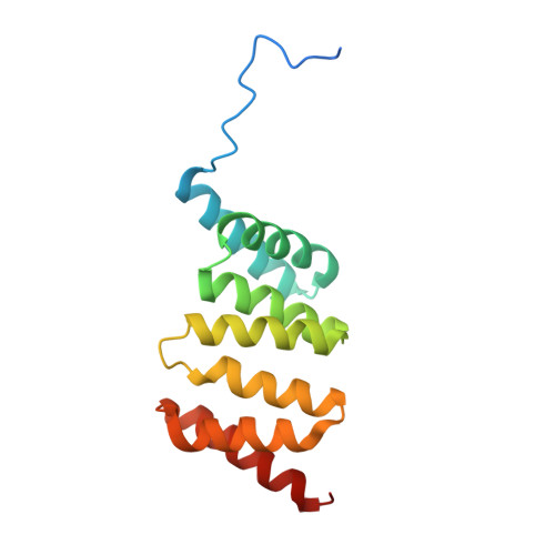
    3KS2

  • PubMed Abstract: 

    Type III secretion systems are a common virulence mechanism in many Gram-negative bacterial pathogens. These systems use a nanomachine resembling a molecular needle and syringe to provide an energized conduit for the translocation of effector proteins from the bacterial cytoplasm to the host cell cytoplasm for the benefit of the pathogen. Prior to translocation specialized chaperones maintain proper effector protein conformation. The class II chaperone, Invasion plasmid gene (Ipg) C, stabilizes two pore forming translocator proteins. IpgC exists as a functional dimer to facilitate the mutually exclusive binding of both translocators.


  • Organizational Affiliation

    Division of Cell Biology and Biophysics, School of Biological Sciences, University of Missouri-Kansas City, Kansas City, MO, USA.


Macromolecules
Find similar proteins by:  (by identity cutoff)  |  3D Structure
Entity ID: 1
MoleculeChains Sequence LengthOrganismDetailsImage
Chaperone protein ipgC
A, B, C, D, E
A, B, C, D, E, F, G, H, I, J, K, L, M, N, O, P, Q, R
151Shigella flexneriMutation(s): 0 
Gene Names: ipgC
UniProt
Find proteins for P0A2U4 (Shigella flexneri)
Explore P0A2U4 
Go to UniProtKB:  P0A2U4
Entity Groups  
Sequence Clusters30% Identity50% Identity70% Identity90% Identity95% Identity100% Identity
UniProt GroupP0A2U4
Sequence Annotations
Expand
  • Reference Sequence
Experimental Data & Validation

Experimental Data

  • Method: X-RAY DIFFRACTION
  • Resolution: 3.30 Å
  • R-Value Free: 0.296 
  • R-Value Work: 0.259 
  • R-Value Observed: 0.261 
  • Space Group: P 1 21 1
Unit Cell:
Length ( Å )Angle ( ˚ )
a = 140.496α = 90
b = 71.474β = 93.86
c = 171.012γ = 90
Software Package:
Software NamePurpose
HKL-2000data collection
PHASERphasing
PHENIXrefinement
HKL-2000data reduction
HKL-2000data scaling

Structure Validation

View Full Validation Report


Entry History 

Deposition Data

Revision History  (Full details and data files)

  • Version 1.0: 2010-08-25
    Type: Initial release
  • Version 1.1: 2011-07-13
    Changes: Version format compliance
  • Version 1.2: 2023-11-01
    Changes: Data collection, Database references, Refinement description