Elimination of a cis-proline-containing loop and turn optimization stabilizes a protein and accelerates its folding.
Jakob, R.P., Zierer, B.K., Weininger, U., Hofmann, S.D., Lorenz, S.H., Balbach, J., Dobbek, H., Schmid, F.X.(2010) J Mol Biol 399: 331-346
- PubMed: 20394751
- DOI: https://doi.org/10.1016/j.jmb.2010.04.007
- Primary Citation of Related Structures:
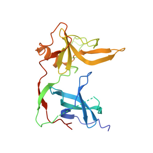
3KNQ - PubMed Abstract:
In the N2 domain of the gene-3-protein of phage fd, two consecutive beta-strands are connected by a mobile loop of seven residues (157-163). The stability of this loop is low, and the Asp160-Pro161 bond at its tip shows conformational heterogeneity with 90% being in the cis and 10% in the trans form. The refolding kinetics of N2 are complex because the molecules with cis or trans isomers at Pro161 both fold to native-like conformations, albeit with different rates. We employed consensus design to shorten the seven-residue irregular loop around Pro161 to a four-residue type I' turn without a proline. This increased the conformational stability of N2 by almost 10 kJ mol(-1) and abolished the complexity of the folding kinetics. Turn sequences obtained from in vitro selections for increased stability strongly resembled those derived from the consensus design. Two other type I' turns of N2 could also be stabilized by consensus design. For all three turns, the gain in stability originates from an increase in the rate of refolding. The turns form native-like structures early during refolding and thus stabilize the folding transition state. The crystal structure of the variant with all three stabilized turns confirms that the 157-163 loop was in fact shortened to a type I' turn and that the other turns maintained their type I' conformation after sequence optimization.
Organizational Affiliation:
Laboratorium für Biochemie and Bayreuther Zentrum für Molekulare Biowissenschaften, Universität Bayreuth, D-95440 Bayreuth, Germany.