3KM9

Structure of complement C5 in complex with the C-terminal beta-grasp domain of SSL7


Experimental Data Snapshot

  • Method: X-RAY DIFFRACTION
  • Resolution: 4.20 Å
  • R-Value Free: 0.297 
  • R-Value Work: 0.233 
  • R-Value Observed: 0.236 

wwPDB Validation   3D Report Full Report


Ligand Structure Quality Assessment 


This is version 2.1 of the entry. See complete history


Literature

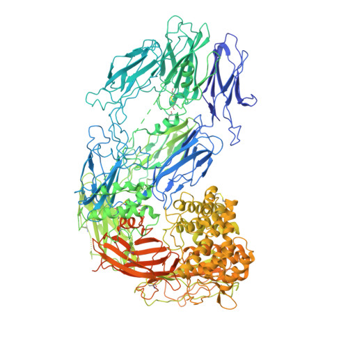
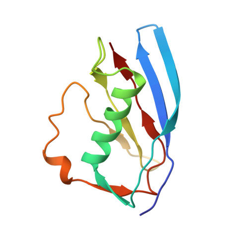
Structural basis for inhibition of complement C5 by the SSL7 protein from Staphylococcus aureus

Laursen, N.S.Gordon, N.Hermans, S.Lorenz, N.Jackson, N.Wines, B.Spillner, E.Christensen, J.B.Jensen, M.Fredslund, F.Bjerre, M.Sottrup-Jensen, L.Fraser, J.D.Andersen, G.R.

(2010) Proc Natl Acad Sci U S A 107: 3681-3686

  • DOI: https://doi.org/10.1073/pnas.0910565107
  • Primary Citation of Related Structures:  
    3KLS, 3KM9

  • PubMed Abstract: 

    Staphylococcus aureus secretes the SSL7 protein as part of its immune evasion strategy. The protein binds both complement C5 and IgA, yet it is unclear whether SSL7 cross-links these two proteins and, if so, what purpose this serves the pathogen. We have isolated a stable IgA-SSL7-C5 complex, and our crystal structure of the C5-SSL7 complex confirms that binding to C5 occurs exclusively through the C-terminal beta-grasp domain of SSL7 leaving the OB domain free to interact with IgA. SSL7 interacts with C5 >70 A from the C5a cleavage site without inducing significant conformational changes in C5, and efficient inhibition of convertase cleavage of C5 is shown to be IgA dependent. Inhibition of C5a production and bacteriolysis are all shown to require C5 and IgA binding while inhibition of hemolysis is achieved by the C5 binding SSL7 beta-grasp domain alone. These results provide a conceptual and structural basis for the development of a highly specific complement inhibitor preventing only the formation of the lytic membrane attack complex without affecting the important signaling functions of C5a.


  • Organizational Affiliation

    Department of Molecular Biology, University of Aarhus, DK-8000 Aarhus, Denmark.


Macromolecules
Find similar proteins by:  (by identity cutoff)  |  3D Structure
Entity ID: 1
MoleculeChains Sequence LengthOrganismDetailsImage
Complement C5A,
C [auth B]
1,676Homo sapiensMutation(s): 0 
UniProt & NIH Common Fund Data Resources
Find proteins for P01031 (Homo sapiens)
Explore P01031 
Go to UniProtKB:  P01031
PHAROS:  P01031
GTEx:  ENSG00000106804 
Entity Groups  
Sequence Clusters30% Identity50% Identity70% Identity90% Identity95% Identity100% Identity
UniProt GroupP01031
Sequence Annotations
Expand
  • Reference Sequence
Find similar proteins by:  (by identity cutoff)  |  3D Structure
Entity ID: 2
MoleculeChains Sequence LengthOrganismDetailsImage
Staphylococcal enterotoxin-like toxinB [auth X],
D [auth Y]
103Staphylococcus aureus subsp. aureus str. NewmanMutation(s): 0 
UniProt
Find proteins for A0A0H3K6Z8 (Staphylococcus aureus (strain Newman))
Explore A0A0H3K6Z8 
Go to UniProtKB:  A0A0H3K6Z8
Entity Groups  
Sequence Clusters30% Identity50% Identity70% Identity90% Identity95% Identity100% Identity
UniProt GroupA0A0H3K6Z8
Sequence Annotations
Expand
  • Reference Sequence
Oligosaccharides

Help

Entity ID: 3
MoleculeChains Length2D Diagram Glycosylation3D Interactions
2-acetamido-2-deoxy-beta-D-glucopyranose-(1-4)-2-acetamido-2-deoxy-beta-D-glucopyranoseE [auth C],
F [auth D]
2N-Glycosylation
Glycosylation Resources
GlyTouCan:  G42666HT
GlyCosmos:  G42666HT
GlyGen:  G42666HT
Experimental Data & Validation

Experimental Data

  • Method: X-RAY DIFFRACTION
  • Resolution: 4.20 Å
  • R-Value Free: 0.297 
  • R-Value Work: 0.233 
  • R-Value Observed: 0.236 
  • Space Group: P 31
Unit Cell:
Length ( Å )Angle ( ˚ )
a = 144.79α = 90
b = 144.79β = 90
c = 245.28γ = 120
Software Package:
Software NamePurpose
PHASERphasing
PHENIXrefinement
XDSdata reduction
XSCALEdata scaling

Structure Validation

View Full Validation Report


Ligand Structure Quality Assessment 


Entry History 

Revision History  (Full details and data files)

  • Version 1.0: 2009-12-01
    Type: Initial release
  • Version 1.1: 2011-07-13
    Changes: Source and taxonomy, Version format compliance
  • Version 1.2: 2017-11-01
    Changes: Refinement description
  • Version 2.0: 2020-07-29
    Type: Remediation
    Reason: Carbohydrate remediation
    Changes: Atomic model, Data collection, Derived calculations, Structure summary
  • Version 2.1: 2023-11-01
    Changes: Data collection, Database references, Refinement description, Structure summary