3K3F

Crystal Structure of the Urea Transporter from Desulfovibrio Vulgaris


Experimental Data Snapshot

  • Method: X-RAY DIFFRACTION
  • Resolution: 2.30 Å
  • R-Value Free: 0.204 
  • R-Value Work: 0.179 
  • R-Value Observed: 0.180 

wwPDB Validation   3D Report Full Report


This is version 1.3 of the entry. See complete history


Literature

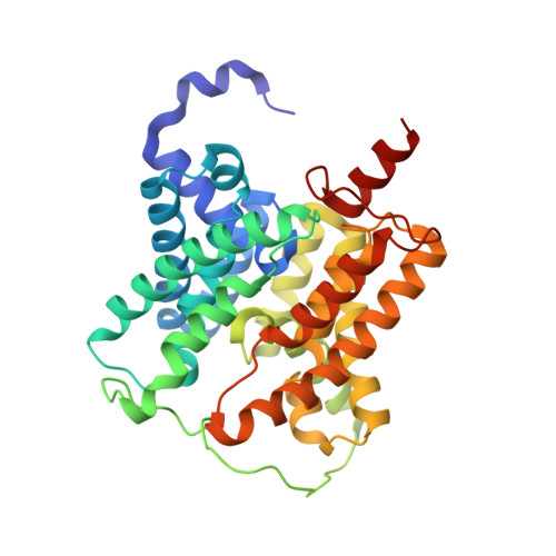
Crystal structure of a bacterial homologue of the kidney urea transporter.

Levin, E.J.Quick, M.Zhou, M.

(2009) Nature 462: 757-761

  • DOI: https://doi.org/10.1038/nature08558
  • Primary Citation of Related Structures:  
    3K3F, 3K3G

  • PubMed Abstract: 

    Urea is highly concentrated in the mammalian kidney to produce the osmotic gradient necessary for water re-absorption. Free diffusion of urea across cell membranes is slow owing to its high polarity, and specialized urea transporters have evolved to achieve rapid and selective urea permeation. Here we present the 2.3 A structure of a functional urea transporter from the bacterium Desulfovibrio vulgaris. The transporter is a homotrimer, and each subunit contains a continuous membrane-spanning pore formed by the two homologous halves of the protein. The pore contains a constricted selectivity filter that can accommodate several dehydrated urea molecules in single file. Backbone and side-chain oxygen atoms provide continuous coordination of urea as it progresses through the filter, and well-placed alpha-helix dipoles provide further compensation for dehydration energy. These results establish that the urea transporter operates by a channel-like mechanism and reveal the physical and chemical basis of urea selectivity.


  • Organizational Affiliation

    Department of Physiology & Cellular Biophysics, College of Physicians and Surgeons, Columbia University, 630 West 168th Street, New York, New York 10032, USA.


Macromolecules
Find similar proteins by:  (by identity cutoff)  |  3D Structure
Entity ID: 1
MoleculeChains Sequence LengthOrganismDetailsImage
Urea transporter340Nitratidesulfovibrio vulgaris str. HildenboroughMutation(s): 0 
Gene Names: DVU_1160
Membrane Entity: Yes 
UniProt
Find proteins for Q72CX3 (Desulfovibrio vulgaris (strain ATCC 29579 / DSM 644 / NCIMB 8303 / VKM B-1760 / Hildenborough))
Explore Q72CX3 
Go to UniProtKB:  Q72CX3
Entity Groups  
Sequence Clusters30% Identity50% Identity70% Identity90% Identity95% Identity100% Identity
UniProt GroupQ72CX3
Sequence Annotations
Expand
  • Reference Sequence
Experimental Data & Validation

Experimental Data

  • Method: X-RAY DIFFRACTION
  • Resolution: 2.30 Å
  • R-Value Free: 0.204 
  • R-Value Work: 0.179 
  • R-Value Observed: 0.180 
  • Space Group: P 63
Unit Cell:
Length ( Å )Angle ( ˚ )
a = 110.131α = 90
b = 110.131β = 90
c = 84.861γ = 120
Software Package:
Software NamePurpose
DENZOdata reduction
SCALEPACKdata scaling
SHELXphasing
REFMACrefinement
PDB_EXTRACTdata extraction
HKL-2000data collection
HKL-2000data reduction
SOLVEphasing

Structure Validation

View Full Validation Report


Entry History 

Deposition Data

Revision History  (Full details and data files)

  • Version 1.0: 2009-11-17
    Type: Initial release
  • Version 1.1: 2011-07-13
    Changes: Advisory, Source and taxonomy, Version format compliance
  • Version 1.2: 2017-11-01
    Changes: Data collection, Refinement description
  • Version 1.3: 2024-02-21
    Changes: Data collection, Database references, Derived calculations