Catalytic reaction mechanism of Pseudomonas stutzeri l-rhamnose isomerase deduced from X-ray structures
Yoshida, H., Yamaji, M., Ishii, T., Izumori, K., Kamitori, S.(2010) FEBS J 277: 1045-1057
- PubMed: 20088877
- DOI: https://doi.org/10.1111/j.1742-4658.2009.07548.x
- Primary Citation of Related Structures:
3ITT, 3ITV, 3ITX, 3ITY, 3IUD, 3IUH, 3IUI - PubMed Abstract:
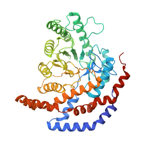
L-Rhamnose isomerase (L-RhI) catalyzes the reversible isomerization of L-rhamnose to L-rhamnulose. Pseudomonas stutzeril-RhI, with a broad substrate specificity, can catalyze not only the isomerization of L-rhamnose, but also that between D-allose and D-psicose. For the aldose-ketose isomerization by L-RhI, a metal-mediated hydride-shift mechanism has been proposed, but the catalytic mechanism is still not entirely understood. To elucidate the entire reaction mechanism, the X-ray structures of P. stutzeril-RhI in an Mn(2+)-bound form, and of two inactive mutant forms of P. stutzeril-RhI (S329K and D327N) in a complex with substrate/product, were determined. The structure of the Mn(2+)-bound enzyme indicated that the catalytic site interconverts between two forms with the displacement of the metal ion to recognize both pyranose and furanose ring substrates. Solving the structures of S329K-substrates allowed us to examine the metal-mediated hydride-shift mechanism of L-RhI in detail. The structural analysis of D327N-substrates and additional modeling revealed Asp327 to be responsible for the ring opening of furanose, and a water molecule coordinating with the metal ion to be involved in the ring opening of pyranose.
Organizational Affiliation:
Life Science Research Center and Faculty of Medicine, Kagawa University, Japan.