3IBY

Structure of cytosolic domain of L. pneumophila FeoB


Experimental Data Snapshot

  • Method: X-RAY DIFFRACTION
  • Resolution: 2.50 Å
  • R-Value Free: 0.288 
  • R-Value Work: 0.212 
  • R-Value Observed: 0.216 

wwPDB Validation   3D Report Full Report


This is version 1.3 of the entry. See complete history


Literature

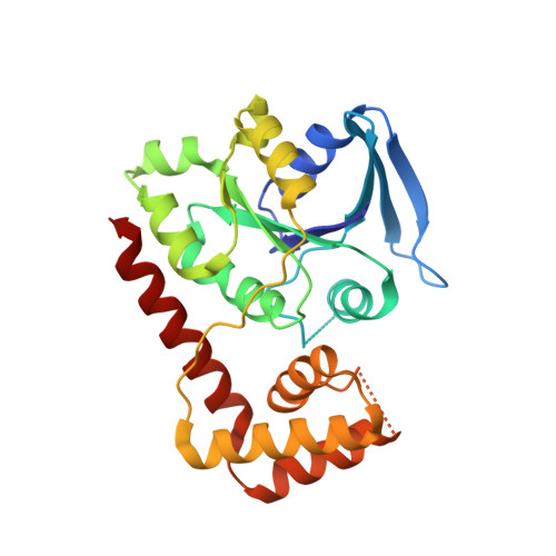
Structure of the GTPase and GDI domains of FeoB, the ferrous iron transporter of Legionella pneumophila.

Petermann, N.Hansen, G.Schmidt, C.L.Hilgenfeld, R.

(2010) FEBS Lett 584: 733-738

  • DOI: https://doi.org/10.1016/j.febslet.2009.12.045
  • Primary Citation of Related Structures:  
    3IBY

  • PubMed Abstract: 

    Prokaryotic pathogens have developed specialized mechanisms for efficient uptake of ferrous iron (Fe(2+)) from the host. In Legionella pneumophila, the causative agent of Legionnaires' disease, the transmembrane GTPase FeoB plays a key role in Fe(2+) acquisition and virulence. FeoB consists of a membrane-embedded core and an N-terminal, cytosolic region (NFeoB). Here, we report the crystal structure of NFeoB from L. pneumophila, revealing a monomeric protein comprising two separate domains with GTPase and guanine-nucleotide dissociation inhibitor (GDI) functions. The GDI domain displays a novel fold, whereas the overall structure of the GTPase domain resembles that of known G domains but is in the rarely observed nucleotide-free state.


  • Organizational Affiliation

    Institute of Biochemistry, Center for Structural and Cell Biology in Medicine, University of Lübeck, Lübeck, Germany.


Macromolecules
Find similar proteins by:  (by identity cutoff)  |  3D Structure
Entity ID: 1
MoleculeChains Sequence LengthOrganismDetailsImage
Ferrous iron transport protein B
A, B, C, D
256Legionella pneumophilaMutation(s): 0 
Gene Names: feoB
UniProt
Find proteins for Q8GNS3 (Legionella pneumophila)
Explore Q8GNS3 
Go to UniProtKB:  Q8GNS3
Entity Groups  
Sequence Clusters30% Identity50% Identity70% Identity90% Identity95% Identity100% Identity
UniProt GroupQ8GNS3
Sequence Annotations
Expand
  • Reference Sequence
Experimental Data & Validation

Experimental Data

  • Method: X-RAY DIFFRACTION
  • Resolution: 2.50 Å
  • R-Value Free: 0.288 
  • R-Value Work: 0.212 
  • R-Value Observed: 0.216 
  • Space Group: P 21 21 21
Unit Cell:
Length ( Å )Angle ( ˚ )
a = 53.056α = 90
b = 130.697β = 90
c = 157.616γ = 90
Software Package:
Software NamePurpose
ProDCdata collection
PHASERphasing
REFMACrefinement
XDSdata reduction
SCALAdata scaling

Structure Validation

View Full Validation Report


Entry History 

Deposition Data

Revision History  (Full details and data files)

  • Version 1.0: 2009-10-13
    Type: Initial release
  • Version 1.1: 2011-07-13
    Changes: Advisory, Version format compliance
  • Version 1.2: 2018-03-07
    Changes: Data collection
  • Version 1.3: 2024-02-21
    Changes: Data collection, Database references